
Radius (bone)
| Radius | |
|---|---|
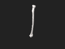
 The radius (shown in red) is a bone in the forearm. | |
| Details | |
| Identifiers | |
| Latin | radius |
| MeSH | D011884 |
| TA98 | A02.4.05.001 |
| TA2 | 1210 |
| FMA | 23463 |
| Anatomical terms of bone | |
The radius or radial bone (pl.: radii or radiuses) is one of the two large
The radius is part of two
The corresponding bone in the lower leg is the tibia.
Structure


The long narrow
The
The radius has a body and two extremities. The


Near the wrist
The distal end of the radius is large and of quadrilateral form.
- Joint surfaces
It is provided with two articular surfaces – one below, for the carpus, and another at the medial side, for the ulna.
- The carpal articular surface is triangular, concave, smooth, and divided by a slight antero-posterior ridge into two parts. Of these, the lateral, triangular, articulates with the scaphoid bone; the medial, quadrilateral, with the lunate bone.
- The articular surface for the ulna is called the ulnar notch(sigmoid cavity) of the radius; it is narrow, concave, smooth, and articulates with the head of the ulna.
These two articular surfaces are separated by a prominent ridge, to which the base of the triangular articular disk is attached; this disk separates the wrist-joint from the distal radioulnar articulation.
- Other surfaces
This end of the bone has three non-articular surfaces – volar, dorsal, and lateral.
- The volar surface, rough and irregular, affords attachment to the volar radiocarpal ligament.
- The dorsal surface is convex, affords attachment to the dorsal radiocarpal ligament, and is marked by three grooves. Enumerated from the lateral side:
- The first groove is broad, but shallow, and subdivided into two by a slight ridge: the lateral of these two, transmits the tendon of the extensor carpi radialis longus muscle; the medial, the tendon of the extensor carpi radialis brevis muscle.
- The second is deep but narrow, and bounded laterally by a sharply defined ridge; it is directed obliquely from above downward and lateralward, and transmits the tendon of the extensor pollicis longus muscle.
- The third is broad, for the passage of the tendons of the extensor digitorum communis.
- The lateral surface is prolonged obliquely downward into a strong, conical projection, the styloid process, which gives attachment by its base to the tendon of the brachioradialis, and by its apex to the extensor pollicis brevismuscle.
Body
The body of the radius (or shaft of radius) is prismoid in form, narrower above than below, and slightly curved, so as to be convex lateralward. It presents three borders and three surfaces.
- Borders
The volar border (margo volaris; anterior border; palmar;) extends from the lower part of the
The dorsal border (margo dorsalis; posterior border) begins above at the back of the neck, and ends below at the posterior part of the base of the styloid process; it separates the posterior from the lateral surface. is indistinct above and below, but well-marked in the middle third of the bone.
The interosseous border (internal border; crista interossea; interosseous crest;) begins above, at the back part of the tuberosity, and its upper part is rounded and indistinct; it becomes sharp and prominent as it descends, and at its lower part divides into two ridges which are continued to the anterior and posterior margins of the ulnar notch. To the posterior of the two ridges the lower part of the interosseous membrane is attached, while the triangular surface between the ridges gives insertion to part of the pronator quadratus muscle. This crest separates the volar from the dorsal surface, and gives attachment to the interosseous membrane. The connection between the two bones is actually a joint referred to as a syndesmosis joint.
- Surfaces
The volar surface (facies volaris; anterior surface) is concave in its upper three-fourths, and gives origin to the flexor pollicis longus muscle; it is broad and flat in its lower fourth, and affords insertion to the Pronator quadratus. A prominent ridge limits the insertion of the Pronator quadratus below, and between this and the inferior border is a triangular rough surface for the attachment of the volar radiocarpal ligament. At the junction of the upper and middle thirds of the volar surface is the nutrient foramen, which is directed obliquely upward.
The dorsal surface (facies dorsalis; posterior surface) is convex, and smooth in the upper third of its extent, and covered by the Supinator. Its middle third is broad, slightly concave, and gives origin to the Abductor pollicis longus above, and the extensor pollicis brevis muscle below. Its lower third is broad, convex, and covered by the tendons of the muscles which subsequently run in the grooves on the lower end of the bone.
The lateral surface (facies lateralis; external surface) is convex throughout its entire extent and is known as the convexity of the radius, curving outwards to be convex at the side. Its upper third gives insertion to the supinator muscle. About its center is a rough ridge, for the insertion of the pronator teres muscle.[2] Its lower part is narrow, and covered by the tendons of the abductor pollicis longus muscle and extensor pollicis brevis muscle.
Near the elbow
The upper extremity of the radius (or proximal extremity) presents a head, neck, and tuberosity.
- The radial head has a cylindrical form, and on its upper surface is a shallow cup or fovea for articulation with the radial notch of the ulna, narrow in the rest of its extent, which is embraced by the annular ligament. The deepest point in the fovea is not axi-symmetric with the long axis of the radius, creating a cam effect during pronation and supination.
- The head is supported on a round, smooth, and constricted portion called the neck, on the back of which is a slight ridge for the insertion of part of the supinator muscle.
- Beneath the neck, on the medial side, is an eminence, the and the bone.
Development
The radius is
Ossification commences in the lower end between 9 and 26 months of age.[citation needed] The ossification center for the upper end appears by the fifth year.
The upper epiphysis fuses with the body at the age of seventeen or eighteen years, the lower about the age of twenty.
An additional center sometimes found in the radial tuberosity, appears about the fourteenth or fifteenth year.
Function
Muscle attachments
The
Clinical significance
Radial aplasia refers to the congenital absence or shortness of the radius.
Fracture

Specific fracture types of the radius include:
- Proximal radius fracture. A fracture within the capsule of the elbow joint results in the fat pad sign or "sail sign" which is a displacement of the fat pad at the elbow.

- interosseous membrane.[3]
- Radial shaft fracture
- Distal radius fracture
- distal radioulnar joint
- Colles' fracture – a distal fracture of the radius with dorsal (posterior) displacement of the wrist and hand
- Smith's fracture – a distal fracture of the radius with volar (ventral) displacement of the wrist and hand
- radiocarpal joint.
History
The word radius is
The radius is named so because the radius (bone) acts like the radius (of a circle). It rotates around the ulna and the far end (where it joins to the bones of the hand), known as the styloid process of the radius, is[clarification needed] the distance from the ulna (center of the circle) to the edge of the radius (the circle). The ulna acts as the center point to the circle because when the arm is rotated the ulna does not move.
Other animals
In four-legged animals, the radius is the main load-bearing bone of the lower forelimb. Its structure is similar in most terrestrial
Gallery
-
 Radius bone and radius of a circle comparison.
 -
 Position of radius (shown in red).
 -
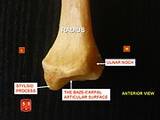 Radius, styloid process - anterior view
 -
 Radius, ulnar notch - posterior view
 -
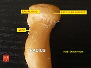 Radius, radial head – posterior view
 -
 Radius, radial head – anterior view
 -
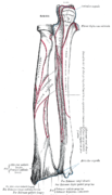
 Radius l. dx. – ant. view
 -
 Radius l. dx. – post. view
 -
 Anterior surface of radius (at right)
 -
 Posterior surface of radius (at left)
 -
 Posterior view of right proximal radius
 -
 Posterior view of right distal radius
 -
 Medial view of right proximal radius
 -
 Medial view of right distal radius
 -
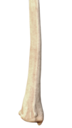
 Lateral view of right distal radius
 -
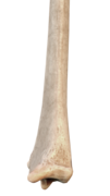
 Anterior view of right distal radius
 -
 Anterior view of right proximal radius
 -
Radius bone anatomy


References
![]() This article incorporates text in the public domain from page 219 of the 20th edition of Gray's Anatomy (1918)
This article incorporates text in the public domain from page 219 of the 20th edition of Gray's Anatomy (1918)
- ^ Clemente, Carmine D. (2007), Anatomy: A Regional Atlas of the Human Body (5th ed.), Philadelphia, PA: Lippincott Williams & Wilkins
- ISBN 978-0-7817-6274-8.
- ^ Essex Lopresti fracture at Wheeless' Textbook of Orthopaedics online
- ^ Marieb, E., R.N., Ph.D; Mallatt, J., Ph.D. & Wilhelm, P., Ph.D. (2008), Human Anatomy (5th ed.), San Francisco, CA: Pearson Benjamin Cummings, p. 188
{{citation}}: CS1 maint: multiple names: authors list (link) - ISBN 0-03-910284-X.