
Renal artery
| Renal artery | |
|---|---|
segmental arteries, Ovarian artery | |
| Vein | Renal vein |
| Supplies | Kidneys |
| Identifiers | |
| Latin | arteria renalis |
| MeSH | D012077 |
| TA98 | A12.2.12.075 |
| TA2 | 4269 |
| FMA | 14751 |
| Anatomical terminology] | |
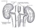
The renal arteries are paired arteries that supply the
The renal arteries carry a large portion of total blood flow to the kidneys. Up to a third of total cardiac output can pass through the renal arteries to be filtered by the kidneys.
Structure
The renal arteries normally arise at a 90° angle off of the left interior side of the abdominal aorta, immediately below the superior mesenteric artery.[1] They have a radius of approximately 0.25 cm,[2] 0.26 cm at the root.[3] The measured mean diameter can differ depending on the imaging method used. For example, the diameter was found to be 5.04 ± 0.74 mm using ultrasound but 5.68 ± 1.19 mm using angiography.[4][5]
Due to the anatomical position of the aorta, the inferior vena cava, and the kidneys, the right renal artery is normally longer than the left renal artery.[1][6]
- The right passes behind the head of the pancreas, and the descending part of the duodenum. It’s somewhat lower than the left one.
- Left artery lies behind the left renal vein, the body of the pancreas and the splenic vein, and is crossed by the inferior mesenteric vein.
Branches
Before reaching the
Each vessel gives off some small
One or two accessory renal arteries are frequently found, especially on the left side since they usually arise from the aorta, and may come off above (more common) or below the main artery. Instead of entering the kidney at the hilus, they usually pierce the upper or lower part of the organ.
Variation
The arterial supply of the kidneys is variable and there may be one or more renal arteries supplying each kidney.[1] It is located above the renal vein. Supernumerary renal arteries (two or more arteries to a single kidney) are the most common renovascular anomaly, occurrence ranging from 25% to 40% of kidneys.[8] Aberrant renal arteries may be present, and may complicate surgical procedures.[9]
Clinical significance
Stenosis
Renal artery stenosis, or narrowing of one or both renal arteries will lead to hypertension as the affected kidneys release renin to increase blood pressure to preserve perfusion to the kidneys. RAS is typically diagnosed with duplex ultrasonography of the renal arteries. It is treated with the use of balloon angioplasty and stents, if necessary.
Atherosclerosis
Renal artery aneurysm
A dilated renal artery measuring twice its normal size indicates a renal artery aneurysm.[4]
Trauma
A renal artery is damaged in 4% of blunt traumas and 7% of penetrating traumas to the abdomen.[10]
Additional images
-
 Volume rendered CT scan of abdominal and pelvic blood vessels.
 -
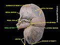
 The posterior surfaces of the kidneys, showing areas of relation to the parietes.
 -
 Renal artery
 -
 Renal artery
 -
 Renal artery
 -
 Renal artery
.gif)


References
![]() This article incorporates text in the public domain from page 610 of the 20th edition of Gray's Anatomy (1918)
This article incorporates text in the public domain from page 610 of the 20th edition of Gray's Anatomy (1918)
- ^ ISBN 978-0-323-67376-1, retrieved 2021-01-13
- PMID 15967872.
- ISBN 978-4-274-90318-2.[page needed]
- ^ a b Renal Artery Aneurysm at eMedicine
- S2CID 24188140.
- .
- PMID 29083626. Retrieved 15 Nov 2021.
- PMID 31950928.
- PMID 4766019.
- ISBN 978-0-8151-4369-7, retrieved 2021-01-13
External links
- MedlinePlus Image 9818
- Anatomy photo:40:11-0105 at the SUNY Downstate Medical Center - "Posterior Abdominal Wall: Branches of the Abdominal Aorta"