
Renal pelvis
| Renal pelvis | |
|---|---|
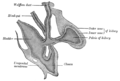
 Cross-section of the kidney, with major structures labelled. The renal pelvis, located in the middle of the image, collects urine from the urinary calices. | |
 An image showing just the pelvis and calices of the kidneys, with the rest of the kidney removed, from a dissected cow and seal specimen. These vary greatly in size and number depending on species.[citation needed] | |
| Details | |
| Precursor | Ureteric bud |
| System | Urinary system |
| Identifiers | |
| Latin | pelvis renalis |
| MeSH | D007682 |
| TA98 | A08.1.05.001 |
| TA2 | 3384 |
| FMA | 15575 |
| Anatomical terminology | |
The renal pelvis or pelvis of the kidney is the funnel-like dilated part of the ureter in the kidney. It is formed by the convergence of the major calyces, acting as a funnel for urine flowing from the major calyces to the ureter. It has a mucous membrane and is covered with transitional epithelium and an underlying lamina propria of loose-to-dense connective tissue.
The renal pelvis is situated within the renal sinus alongside the other structures of the renal sinus.[1]
Clinical significance
The renal pelvis is the location of several kinds of
staghorn
" kidney stone may block all or part of the renal pelvis.
The size of the renal pelvis plays a major role in the
gestational age and 7 mm afterwards.[2] In adults, 13% of the normal population have a transverse pelvic diameter of over 10 mm.[3]
Etymology and pronunciation
Like the
combining form pyelo- denotes the renal pelvis (pyelo- is not to be confused with pyo-). The words infundibulum and choana are other words for funnel-shaped cavities (which medical English got from the Latin and Greek words for "funnel", respectively), and the renal pelvis is sometimes called the renal infundibulum. The form *renal choana is logical but is not used
.
Additional images
-
 Depiction of the developing renal pelvis.
 -
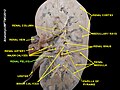 Cutaway section of a preserved human kidney


See also
- Renal sinus
- Pyelectasis, the dilation of the renal pelvis
References
- OCLC 1132300315.)
{{cite book}}: CS1 maint: location missing publisher (link) CS1 maint: others (link - ISBN 9789351523376.
- PMID 8369185.
- ^ Merriam-Webster, Merriam-Webster's Unabridged Dictionary, Merriam-Webster, archived from the original on 2020-05-25, retrieved 2016-02-08.
External links
- Anatomy figure: 40:03-07 at Human Anatomy Online, SUNY Downstate Medical Center—"Section of the kidney, anterior view."
- Anatomy image:8962 at the SUNY Downstate Medical Center
- Anatomy photo: Urinary/mammal/pelvis0/pelvis1 - Comparative Organology at University of California, Davis—"Mammal, renal pelvis (Gross, Medium)"
- Anatomy photo: Urinary/mammal/pelvis1/pelvis1 - Comparative Organology at University of California, Davis—"Mammal, renal pelvis (LM, Medium)"
| National | |
|---|---|
| Other | |