
Renal vein
| Renal vein | |
|---|---|
Renal papilla | |
| Details | |
| Drains from | kidney |
| Source | interlobar veins, Left ovarian vein |
| Drains to | inferior vena cava |
| Artery | Renal artery |
| Identifiers | |
| Latin | venae renales |
| MeSH | D012082 |
| TA98 | A12.3.09.009 |
| TA2 | 5000, 5006 |
| FMA | 14334 |
| Anatomical terminology] | |
The renal veins in the renal circulation, are large-calibre[1] veins that drain blood filtered by the kidneys into the inferior vena cava. There is one renal vein draining each kidney.[citation needed] Each renal vein is formed by the convergence of the interlobar veins of one kidney.[2]
Because the inferior vena cava is on the right half of the body, the left renal vein is longer than the right one.
Structure
One renal vein drains each kidney.[citation needed] A renal vein is situated anterior to its corresponding accompanying renal artery. The renal veins empty into the inferior vena cava, entering it at nearly a 90° angle.[1]
Due to the right-ward displacement of the inferior vena cava from the midline, the left renal vein is some 3 times longer than the right one (~7.5 cm and ~2.5 cm, respectively).[1]
The renal vein divides into 4 divisions upon entering the kidney:[contradictory][citation needed]
- the anterior branch which receives blood from the anterior portion of the kidney and,
- the posterior branch which receives blood from the posterior portion.
Tributaries
Because the tributaries of the
Relations
The anatomical relations of the two renal veins are bilaterally asymmetrical.
Left renal vein
The left renal vein is situated posterior to the
Right renal vein
The right renal vein is situated posterior to the descending part of the duodenum.[1]
Variation
There is typically a single renal vein drainin each kidney, but accessory renal veins are commonly encountered; renal vasculature anomalies are more frequent with ectopic kidneys, and almost always present with horseshoe kidney).[4]
In some individuals, the left renal vein passes posterior to the abdominal aorta instead of in anterior to it;[1] this is termed a retro-aortic left renal vein (also known as "The Vein of Schnitker"). If there is both a vein passing in front of and one behind the aorta this is called a circumaortic renal vein. In the case of a left sided IVC and the right renal vein passes behind the abdominal aorta, this is termed a retroaortic right renal vein, which is also known as “The Reverse Vein of Schnitker”.[citation needed]
Clinical significance
Diseases associated with the renal vein include renal vein thrombosis (RVT) and nutcracker syndrome (renal vein entrapment syndrome).[citation needed]
Additional images
-
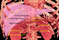 3D-renderedcomputed tomography, showing one renal vein (in red color) for each kidney
 -
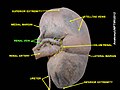
 Frontal section through the kidney
 -
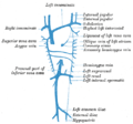 Diagram showing completion of development of the parietal veins.
 -
 The venæ cavæ and azygos veins, with their tributaries.
 -
 Renal vein
 -
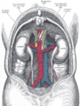 Human kidneys viewed from behind with spine removed.
 -
 Kidney
 -
 Renal vein
 -
 Renal vein
 -
 Renal vein


See also
- Renal physiology
- Nutcracker syndrome
- Renal artery
References
- ^ OCLC 1201341621.)
{{cite book}}: CS1 maint: location missing publisher (link) CS1 maint: others (link - ISBN 9780134320762.
- ^ a b Dissector Answers - Kidney & Retroperitoneum Archived 2007-11-09 at the Wayback Machine
- ^ "Multiple renal veins". Medcyclopaedia. GE. Archived from the original on 2012-02-05.
External links
- Anatomy figure: 40:06-05 at Human Anatomy Online, SUNY Downstate Medical Center - "Retroperitoneal structures on the posterior abdominal wall."