
Rib
This article needs additional citations for verification. (June 2008) |
| Rib | |
|---|---|
 Collection of single ribs in the Faculty of Education of Charles University | |
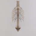
 Animation of all ribs, including the false ones in humans | |
| Details | |
| Identifiers | |
| Latin | costae |
| MeSH | D012272 |
| TA98 | A02.3.01.001 A02.3.02.001 |
| TA2 | 1105, 1118 |
| FMA | 7574 |
| Anatomical terminology | |
In
Human anatomy
Rib details
Human ribs are flat bones that form part of the rib cage to help protect internal organs. Humans usually have 24 ribs, in 12 pairs.[2] 1 in 500 people have an extra rib known as a cervical rib. People may have a cervical rib on the right, left or both sides.[3] All are attached at the back to the thoracic vertebrae and are numbered from 1 to 12 according to the vertebrae to which they attach. The first rib is attached to thoracic vertebra 1 (T1). At the front of the body, most of the ribs are joined by costal cartilage to the sternum. Ribs connect to vertebrae at the costovertebral joints.[4]
The parts of a rib includes the head, neck, body (or shaft), tubercle, and angle.
The head of the rib lies next to a vertebra. The ribs connect to the vertebrae with two costovertebral joints, one on the head and one on the neck. The head of the rib has a superior and an inferior articulating region, separated by a crest. These articulate with the superior and inferior costal facets on the connecting vertebrae.[5] The crest gives attachment to the intra-articulate ligament that joins the rib to the vertebra of the same number, at the intervertebral disc. Another ligament, the radiate ligament joins the head of the rib to both the body of the upper vertebra and to the body of the lower vertebra. The smaller middle part of the ligament connects to the intervertebral disc. This plane joint is known as the articulation of the head of the rib.
The other costovertebral joint is that between the tubercle on the neck and the transverse process of the joining thoracic vertebra of the same rib number, and this is known as the costotransverse joint. The superior costotransverse ligament attaches from the non-articular facet of the tubercle to the transverse process of the vertebra.
The neck of the rib is a flattened part that extends laterally from the head. The neck is about 3 cm long. Its anterior surface is flat and smooth, whilst its posterior is perforated by numerous foramina and its surface rough, to give attachment to the ligament of the neck. Its upper border presents a rough crest (crista colli costae) for the attachment of the anterior costotransverse ligament; its lower border is rounded.
A tubercle of rib on the posterior surface of the neck of the rib, has two facets (surfaces) one articulating and one non-articulating. The articular facet, is small and oval and is the lower and more medial of the two, and connects to the transverse costal facet on the thoracic vertebra of the same rib number.[5] The transverse costal facet is on the end of the transverse process of the lower of the two vertebrae to which the head is connected. The non-articular portion is a rough elevation and affords attachment to the ligament of the tubercle. The tubercle is much more prominent in the upper ribs than in the lower ribs.
Rib cage

The first seven sets of ribs, known as "
In general, human ribs increase in length from ribs 1 through 7 and decrease in length again through rib 12. Along with this change in size, the ribs become progressively oblique (slanted) from ribs 1 through 9, then less slanted through rib 12.[7]
The rib cage is separated from the lower abdomen by the thoracic diaphragm which controls breathing. When the diaphragm contracts, the thoracic cavity is expanded, reducing intra-thoracic pressure and drawing air into the lungs. This happens through one of two actions (or a mix of the two): when the lower ribs the diaphragm connects to are stabilized by muscles and the central tendon is mobile, when the muscle contracts the central tendon is drawn down, compressing the cavity underneath and expanding the thoracic cavity downward. When the central tendon is stabilized and the lower ribs are mobile, a contraction of the diaphragm elevates the ribs, which works in conjunction with other muscles to expand the thoracic indent upward.
Development
Early in
During the fourth week (
The ribs begin as cartilage that later ossifies – a process called endochondral ossification. Primary ossification centers are located near the angle of each rib, and ossification continues in the direction away from the head and neck. During adolescence secondary ossification centers are formed in the tubercles and heads of the ribs.[8]
Other animals


In
In most true tetrapods, many of these early ribs have been lost, and in living
In birds, ribs are present as distinct bones only on the thoracic region, although small fused ribs are present on the
Ribs as food
.Animated images
-
 Thoracic cage with spine

See also
References
- ISBN 978-0-323-17281-3, retrieved 2020-11-03
- ^ ISBN 978-0-323-04048-8, retrieved 2020-11-03
- S2CID 256899032.
- ISBN 9781496347213.
- ^ ISBN 9781455704187.
- ISBN 978-0-323-04048-8, retrieved 2020-11-03
- ^ a b Saladin, K. S. (2010). Anatomy and Physiology: The Unity of Form and Function (5th ed.). New York, NY: McGraw-Hill.
- ^ ISBN 0443065837.
- ^ ISBN 0-03-910284-X.