
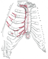
Rib cage
| Rib cage | |
|---|---|
 Human rib cage | |
 Animation of the rib cage | |
| Details | |
| Identifiers | |
| Latin | cavea thoracis |
| MeSH | D000070602 |
| TA98 | A02.3.04.001 |
| TA2 | 1096 |
| FMA | 7480 |
| Anatomical terminology | |
The rib cage or thoracic cage, is an
A typical
In
Structure
There are thirty-three vertebrae in the human vertebral column. The rib cage is associated with TH1−TH12. Ribs are described based on their location and connection with the sternum. All ribs are attached posteriorly to the
Attachment
The terms true ribs and false ribs describe rib pairs that are directly or indirectly attached to the
The phrase floating rib (
The spaces between the ribs are known as
-
 Human rib cage - CT scan (parallel projection (left) and perspective projection (right))
 -
 true / fixed ribsfalse ribsfalse and floating ribs


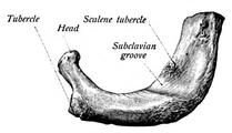
Parts of rib

Each rib consists of a head, neck, and a shaft. All ribs are attached posteriorly to the
The neck of the rib is the flattened part that extends laterally from the head. The neck is about 3 cm long. Its anterior surface is flat and smooth, whilst its posterior is perforated by numerous foramina and its surface rough, to give attachment to the ligament of the neck. Its upper border presents a rough crest (crista colli costae) for the attachment of the anterior costotransverse ligament; its lower border is rounded.
On the posterior surface at the neck, is an eminence—the tubercle that consists of an articular and a non-articular portion. The articular portion is the lower and more medial of the two and presents a small, oval surface for articulation with the transverse costal facet on the end of the transverse process of the lower of the two vertebrae to which the head is connected. The non-articular portion is a rough elevation and affords attachment to the ligament of the tubercle. The tubercle is much more prominent in the upper ribs than in the lower ribs.
The angle of a
The distance between the angle and the tubercle is progressively greater from the second to the tenth ribs. The area between the angle and the tubercle is rounded, rough, and irregular, and serves for the attachment of the
Bones
Ribs and vertebrae
The first rib (the topmost one) is the most curved and usually the shortest of all the ribs; it is broad and flat, its surfaces looking upward and downward, and its borders inward and outward.
-
 First rib seen from above
 -
 Costal groove position on a central rib


The head is small and rounded, and possesses only a single articular facet, for articulation with the body of the first
The second rib is the second uppermost rib in humans or second most frontal in animals that walk on four limbs. In humans, the second rib is defined as a true rib since it connects with the sternum through the intervention of the
The ninth rib has a frontal part at the same level as the
The tenth rib attaches directly to the body of vertebra T10 instead of between vertebrae like the second through ninth ribs. Due to this direct attachment, vertebra T10 has a complete costal facet on its body.[3]

The eleventh and twelfth ribs, the floating ribs, have a single
Sternum
The sternum is a long, flat bone that forms the front of the rib cage. The cartilages of the top seven ribs (the true ribs) join with the sternum at the sternocostal joints. The costal cartilage of the second rib articulates with the sternum at the sternal angle making it easy to locate.[9]
The manubrium is the wider, superior portion of the sternum. The top of the manubrium has a shallow, U-shaped border called the jugular (suprasternal) notch. The clavicular notch is the shallow depression located on either side at the superior-lateral margins of the manubrium. This is the site of the sternoclavicular joint, between the sternum and clavicle. The first ribs also attach to the manubrium.[10]
The
Development
Expansion of the rib cage in males is caused by the effects of testosterone during puberty.[12] Thus, males generally have broad shoulders and expanded chests, allowing them to inhale more air to supply their muscles with oxygen.

Variation
Variations in the number of ribs occur. About 1 in 200–500 people have an additional
In several ethnic groups, most significantly the Japanese, the tenth rib is sometimes a
Function

The human rib cage is a component of the human respiratory system. It encloses the thoracic cavity, which contains the lungs. An inhalation is accomplished when the muscular diaphragm, at the floor of the thoracic cavity, contracts and flattens, while the contraction of intercostal muscles lift the rib cage up and out.
Expansion of the thoracic cavity is driven in three planes; the vertical, the anteroposterior and the transverse. The vertical plane is extended by the help of the diaphragm contracting and the abdominal muscles relaxing to accommodate the downward pressure that is supplied to the abdominal viscera by the diaphragm contracting. A greater extension can be achieved by the diaphragm itself moving down, rather than simply the domes flattening. The second plane is the anteroposterior and this is expanded by a movement known as the 'pump handle'. The downward sloping nature of the upper ribs are as such because they enable this to occur. When the external intercostal muscles contract and lift the ribs, the upper ribs are able also to push the sternum up and out. This movement increases the anteroposterior diameter of the thoracic cavity, and hence aids breathing further. The third, transverse, plane is primarily expanded by the lower ribs (some say it is the 7th to 10th ribs in particular), with the diaphragm's central tendon acting as a fixed point. When the diaphragm contracts, the ribs are able to evert (meaning turn outwards or inside out) and produce what is known as the bucket handle movement, facilitated by gliding at the costovertebral joints. In this way, the transverse diameter is expanded and the lungs can fill.
The circumference of the normal adult human rib cage expands by 3 to 5 cm during inhalation.[15]
Clinical significance
Rib fractures are the most common injury to the rib cage. These most frequently affect the middle ribs. When several adjacent ribs incur two or more fractures each, this can result in a flail chest which is a life-threatening condition.
A dislocated rib can be painful and can be caused simply by coughing, or for example by trauma or lifting heavy weights.[16]
One or more costal cartilages can become inflamed – a condition known as costochondritis; the resulting pain is similar to that of a heart attack.
Abnormalities of the rib cage include pectus excavatum ("sunken chest") and pectus carinatum ("pigeon chest"). A bifid rib is a bifurcated rib, split towards the sternal end, and usually just affecting one of the ribs of a pair. It is a congenital defect affecting about 1.2% of the population. It is often without symptoms though respiratory difficulties and other problems can arise.
Rib removal is the surgical removal of one or more ribs for therapeutic or cosmetic reasons.
Rib resection is the removal of part of a rib.
Regeneration
Since the early part of the 20th century, the ability of the human rib to regenerate itself has been appreciated.[2][5] However, scientific reports demonstrating repair have been sporadic and anecdotal. Currently, this phenomenon is best taken advantage of by craniomaxillofacial surgeons, who use both cartilage and bone material from the rib for jaw, face, and ear reconstruction.[6][8]
The perichondrium is a fibrous sheath of vascular connective tissue surrounding the rib cartilage, containing a source of progenitor stem cells required for rib regeneration.[1]
Society and culture
The position of ribs can be permanently altered by a form of body modification called tightlacing, which uses a corset to compress and move the ribs.
The ribs, particularly their sternal ends, are used as a way of estimating age in forensic pathology due to their progressive ossification.[17]
Biblical Story
The number of ribs as 24 (12 pairs) was noted by the
Other animals

In herpetology, costal grooves refer to lateral indents along the integument of salamanders. The grooves run between the axilla to the groin. Each groove overlies the myotomal septa to mark the position of the internal rib.[20][21]
Additional images
-
 Anterior surface of sternum and costal cartilages
 -
 X-ray image of a human chest, with ribs labelled
 -
 3D model of rib cage
 -
 Surface projections of the trunk, including each rib, and the costal margin


See also
- Articulation of head of rib
- Rachitic rosary
- Terms for anatomical location
- Terms for bones
Notes
![]() This article incorporates text in the public domain from the 20th edition of Gray's Anatomy (1918)
This article incorporates text in the public domain from the 20th edition of Gray's Anatomy (1918)
- ^ a b "The Thoracic Cage · Anatomy and Physiology". Retrieved 10 March 2018.
- ^ ISBN 9780226870137. Retrieved 10 March 2018.
- ^ ISBN 978-0-07-352569-3.
- ^ a b Smith, Sarah. "Intercostal spaces | Radiology Reference Article | Radiopaedia.org". radiopaedia.org.
- ^ a b http://www.teachmeanatomy.com/osteology-of-the-thorax/[permanent dead link]
- ^ ISBN 978-0-7817-7525-0
- ^ Bålens ytanatomi (surface anatomy). Godfried Roomans, Mats Hjortberg and Anca Dragomir. Institution for Anatomy, Uppsala. 2008.
- ^ PMID 32883133.
- ISBN 978-0-7817-7055-2.
- ISBN 978-1-947172-04-3.
- ISBN 978-0-7817-7055-2.
- ^ a b Testosterone causes expansion of rib cage during puberty as one of secondary sex characteristics."Archived copy". Archived from the original on 2015-09-11. Retrieved 2013-12-31.
{{cite web}}: CS1 maint: archived copy as title (link) - ^ PMID 9925395. Retrieved August 13, 2009.
- PMID 16755094.
- ^ Respiratory system examination Archived 2012-03-23 at the Wayback Machine citing: Health & Physical Assessment, Mosby-Year Book, inc. School of Nursing, Peking University, 2003
- ^ "Anatomy of the Human ribs - Dislocated Rib". Dislocated Rib. 2 February 2016. Archived from the original on 12 August 2016.
- PMID 19853490.
- ^ "Chapter 19 On the Bones of the Thorax". Archived from the original on 2007-07-06. Retrieved 2007-08-23.
- ^ Dresden, Danielle (2020-03-12). "How many ribs do humans have? Men, women, and anatomy". Medical News Today. Retrieved 2022-06-05.
Although many people might think that males have fewer ribs than females — most likely sparked by the biblical story of Adam and Eve — there is no factual evidence.
- ISBN 0-8018-4780-X, 9780801847806
- ISBN 978-1-56098-828-1
- ISBN 0-697-21991-7.
References
- Orientation of the intercostal muscle fibers in the human rib cage, Subit D., Glacet A., Hamzah M., Crandall J., Computer Methods in Biomechanics and Biomedical Engineering, 2015, 18, pp. 2064–2065
- Clinically Oriented Anatomy, 4th ed. Keith L. Moore and Robert F. Dalley. pp. 62–64
- Principles of Anatomy Physiology, Tortora GJ and Derrickson B. 11th ED. John Wiley and Sons, 2006. ISBN 0-471-68934-3
- De Humani Corporis Fabrica: online English translation of Vesalius' books on human anatomy.