Serum protein electrophoresis
| Serum protein electrophoresis | |
|---|---|
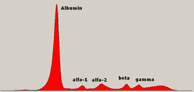
 Normal serum protein electrophoresis diagram with legend of different zones. | |
| MeSH | D001797 |
Serum protein electrophoresis (SPEP or SPE) is a laboratory test that examines specific
Acetate or gel electrophoresis
Proteins are separated by both electrical forces and electroendoosmostic forces. The net charge on a protein is based on the sum charge of its amino acids, and the pH of the buffer. Proteins are applied to a solid matrix such as an agarose gel, or a cellulose acetate membrane in a liquid buffer, and electric current is applied. Proteins with a negative charge will migrate towards the positively charged anode. Albumin has the most negative charge, and will migrate furthest towards the anode. Endoosmotic flow is the movement of liquid towards the cathode, which causes proteins with a weaker charge to move backwards from the application site. Gamma proteins are primarily separated by endoosmotic forces.[4] The drawing of the electrophoretic bands provided by the laboratory may be difficult to remember, and medical students, residents, nurses, and non-specialized medical practitioners may find visual mnemonics useful to recall the five main bands and the shape of normal serum electrophoresis.[5]

Capillary electrophoresis
In capillary electrophoresis, there is no solid matrix. Proteins are separated primarily by strong electroendosmotic forces. The sample is injected into a capillary with a negative surface charge. A high current is applied, and negatively charged proteins such as albumin try to move towards the anode. Liquid buffer flows towards the cathode, and drags proteins with a weaker charge.[6][7]
Serum protein fractions

Albumin
Absence of albumin, known as analbuminaemia, is rare. A decreased level of albumin, however, is common in many diseases, including liver disease, malnutrition, malabsorption, protein-losing nephropathy and enteropathy.[9]
Albumin – alpha-1 interzone
Even staining in this zone is due to alpha-1 lipoprotein (
The high levels of AFP that may occur in hepatocellular carcinoma may result in a sharp band between the albumin and the alpha-1 zone.[citation needed]
Alpha-1 zone
Bence Jones protein may bind to and retard the alpha-1 band. [citation needed]
Alpha-1 – alpha-2 interzone
Two faint bands may be seen representing
Alpha-2 zone
This zone consists principally of
Haptoglobin/haemoglobin complexes migrate more cathodally than haptoglobin as seen in the alpha-2 – beta interzone. This is typically seen as a broadening of the alpha-2 zone.
Alpha-2 macroglobulin may be elevated in children and the elderly. This is seen as a sharp front to the alpha-2 band. AMG is markedly raised (10-fold increase or greater) in association with glomerular protein loss, as in nephrotic syndrome. Due to its large size, AMG cannot pass through glomeruli, while other lower-molecular weight proteins are lost. Enhanced synthesis of AMG accounts for its absolute increase in nephrotic syndrome. Increased AMG is also noted in rats with no albumin indicating that this is a response to low albumin rather than nephrotic syndrome itself[11]
AMG is mildly elevated early in the course of diabetic nephropathy.[citation needed]
Alpha-2 - beta interzone
Cold insoluble globulin forms a band here which is not seen in plasma because it is precipitated by heparin. There are low levels in inflammation and high levels in pregnancy.[citation needed]
Beta lipoprotein forms an irregular
Beta zone
Beta-2 comprises C3 (
Beta-gamma interzone
Gamma zone
The
Hypogammaglobulinaemia is easily identifiable as a "slump" or decrease in the gamma zone. It is normal in infants. It is found in patients with X-linked agammaglobulinemia. IgA deficiency occurs in 1:500 of the population, as is suggested by a pallor in the gamma zone. Of note, hypogammaglobulinema may be seen in the context of MGUS or multiple myeloma.[citation needed]
If the gamma zone shows an increase the first step in interpretation is to establish if the region is narrow or wide. A broad "swell-like" manner (wide) indicates polyclonal immunoglobulin production. If it is elevated in an asymmetric manner or with one or more peaks or narrow "spikes" it could indicate clonal production of one or more immunoglobulins,[13]
Polyclonal gammopathy is indicated by a "swell-like" elevation in the gamma zone, which typically indicates a non-neoplastic condition (although is not exclusive to non-neoplastic conditions). The most common causes of polyclonal hypergammaglobulinaemia detected by electrophoresis are severe infection, chronic liver disease, rheumatoid arthritis, systemic lupus erythematosus and other connective tissue diseases.[citation needed]
A narrow spike is suggestive of a monoclonal gammopathy, also known as a restricted band, or "M-spike". To confirm that the restricted band is an immunoglobulin, follow up testing with
Oligoclonal gammopathy is indicated by one or more discrete clones.[citation needed]
References
- PMID 21374283.
- ISBN 978-1-933864-75-4.
- PMID 4158264.
- ^ Harris 2012, pp. 9–16.
- ^ Medina-De la Garza CE, García-Hernández M, Castro-Corona MA. Visual mnemonics for serum protein electrophoresis https://www.tandfonline.com/doi/full/10.3402/meo.v18i0.22585
- ^ Harris, 2012 & pages 117–123.
- ISBN 0340-81213-3.
- PMID 10437648.
- ^ Peralta, Ruben; Rubery, Brad A (July 30, 2012). Pinsky, Michael R; Sharma, Sat; Talavera, Francisco; Manning, Harold L; Rice, Timothy D (eds.). "Hypoalbuminemia". Medscape. Retrieved 2 October 2013.
- PMID 19870946.
- PMID 9453001.
- ^ Keren 2003, pp. 93–97.
- ^ Tuazon, Sherilyn Alvaran; Scarpaci, Anthony P (May 11, 2012). Staros, Eric B (ed.). "Serum protein electrophoresis". Medscape. Retrieved 2 October 2013.
- PMID 27028734.
- ^ Harris 2012, p. 60.
- PMID 20713974.
External links
- Protein electrophoresis at Lab Tests Online
- Visual mnemonics for serum protein electrophoresis
[1]
