
Skull
| Skull | |
|---|---|
Skeletal system | |
| Identifiers | |
| MeSH | D012886 |
| FMA | 54964 |
| Anatomical terminology] | |
The skull is a
Functions of the skull include protection of the brain, fixing the distance between the eyes to allow
The English word skull is probably derived from
The skull is made up of a number of fused
Structure
Humans


.svg)
The human skull is the bone structure that forms the head in the human skeleton. It supports the structures of the face and forms a cavity for the brain. Like the skulls of other vertebrates, it protects the brain from injury.[5]
The skull consists of three parts, of different embryological origin—the neurocranium, the sutures, and the facial skeleton. The neurocranium (or braincase) forms the protective cranial cavity that surrounds and houses the brain and brainstem.[6] The upper areas of the cranial bones form the calvaria (skullcap). The membranous viscerocranium includes the mandible.
The sutures are fairly rigid joints between bones of the neurocranium.
The facial skeleton is formed by the bones supporting the face.
Bones
Except for the mandible, all of the bones of the skull are joined by
The human skull is generally considered to consist of 22 bones—eight cranial bones and fourteen facial skeleton bones. In the neurocranium these are the occipital bone, two temporal bones, two parietal bones, the sphenoid, ethmoid and frontal bones.
The bones of the facial skeleton (14) are the vomer, two inferior nasal conchae, two nasal bones, two maxilla, the mandible, two palatine bones, two zygomatic bones, and two lacrimal bones. Some sources count a paired bone as one, or the maxilla as having two bones (as its parts); some sources include the hyoid bone or the three ossicles of the middle ear, the malleus, incus, and stapes, but the overall general consensus of the number of bones in the human skull is the stated twenty-two.
Some of these bones—the occipital, parietal, frontal, in the neurocranium, and the nasal, lacrimal, and vomer, in the facial skeleton are flat bones.
Cavities and foramina

The skull also contains
The foramina are openings in the skull. The largest of these is the foramen magnum, of the occipital bone, that allows the passage of the spinal cord as well as nerves and blood vessels.
Processes
The many
Other vertebrates
Fenestrae

|
The fenestrae (from Latin, meaning windows) are openings in the skull.
|
Bones
The
The prefrontal bone is a bone separating the lacrimal and frontal bones in many tetrapod skulls.
Fish

The skull of fishes is formed from a series of only loosely connected bones.
The simpler structure is found in

In
Although the skulls of fossil lobe-finned fish resemble those of the early tetrapods, the same cannot be said of those of the living lungfishes. The skull roof is not fully formed, and consists of multiple, somewhat irregularly shaped bones with no direct relationship to those of tetrapods. The upper jaw is formed from the pterygoids and vomers alone, all of which bear teeth. Much of the skull is formed from cartilage, and its overall structure is reduced.[9]
Tetrapods
The skulls of the earliest
In living tetrapods, a great many of the original bones have either disappeared or fused into one another in various arrangements.
Birds

Birds have a diapsid skull, as in reptiles, with a prelacrimal fossa (present in some reptiles). The skull has a single occipital condyle.[10] The skull consists of five major bones: the frontal (top of head), parietal (back of head), premaxillary and nasal (top beak), and the mandible (bottom beak). The skull of a normal bird usually weighs about 1% of the bird's total bodyweight. The eye occupies a considerable amount of the skull and is surrounded by a sclerotic eye-ring, a ring of tiny bones. This characteristic is also seen in reptiles.
Amphibians

Living amphibians typically have greatly reduced skulls, with many of the bones either absent or wholly or partly replaced by cartilage.[9] In mammals and birds, in particular, modifications of the skull occurred to allow for the expansion of the brain. The fusion between the various bones is especially notable in birds, in which the individual structures may be difficult to identify.
Development

The skull is a complex structure; its bones are formed both by intramembranous and endochondral ossification. The skull roof bones, comprising the bones of the facial skeleton and the sides and roof of the neurocranium, are dermal bones formed by intramembranous ossification, though the temporal bones are formed by endochondral ossification. The endocranium, the bones supporting the brain (the occipital, sphenoid, and ethmoid) are largely formed by endochondral ossification. Thus frontal and parietal bones are purely membranous.[11] The geometry of the skull base and its fossae, the anterior, middle and posterior cranial fossae changes rapidly. The anterior cranial fossa changes especially during the first trimester of pregnancy and skull defects can often develop during this time.[12]
At birth, the human skull is made up of 44 separate bony elements. During development, many of these bony elements gradually fuse together into solid bone (for example, the
The skull in the
Clinical significance
A copper beaten skull is a phenomenon wherein intense intracranial pressure disfigures the internal surface of the skull.[17] The name comes from the fact that the inner skull has the appearance of having been beaten with a ball-peen hammer, such as is often used by coppersmiths. The condition is most common in children.
Injuries and treatment
Injuries to the brain can be life-threatening. Normally the skull protects the brain from damage through its high resistance to deformation; the skull is one of the least deformable structures found in nature, needing the force of about 1 ton to reduce its diameter by 1 cm.
Dating back to
In March 2013, for the first time in the U.S., researchers replaced a large percentage of a patient's skull with a precision, 3D-printed polymer implant.[20] About 9 months later, the first complete cranium replacement with a 3D-printed plastic insert was performed on a Dutch woman. She had been suffering from hyperostosis, which increased the thickness of her skull and compressed her brain.[21]
A study conducted in 2018 by the researchers of Harvard Medical School in Boston, funded by National Institutes of Health (NIH), suggested that instead of travelling via blood, there are "tiny channels" in the skull through which the immune cells combined with the bone marrow reach the areas of inflammation after an injury to the brain tissues.[22]
Transgender procedures
Surgical alteration of sexually dimorphic skull features may be carried out as a part of facial feminization surgery, a set of reconstructive surgical procedures that can alter male facial features to bring them closer in shape and size to typical female facial features.[23][24] These procedures can be an important part of the treatment of transgender people for gender dysphoria.[25][26]
Society and culture
Artificial cranial deformation is a largely historical practice of some cultures. Cords and wooden boards would be used to apply pressure to an infant's skull and alter its shape, sometimes quite significantly. This procedure would begin just after birth and would be carried on for several years.[citation needed]
Osteology
Like the face, the skull and teeth can also indicate a person's life history and origin.
The German physician Franz Joseph Gall in around 1800 formulated the theory of phrenology, which attempted to show that specific features of the skull are associated with certain personality traits or intellectual capabilities of its owner. His theory is now considered to be pseudoscientific.[citation needed]
Sexual dimorphism
In the mid-nineteenth century,
Research has shown that while in early life there is little difference between male and female skulls, in adulthood male skulls tend to be larger and more robust than female skulls, which are lighter and smaller, with a cranial capacity about 10 percent less than that of the male.[28] However, later studies show that women's skulls are slightly thicker and thus men may be more susceptible to head injury than women.[29] However, other studies shows that men's skulls are slightly thicker in certain areas.[30] As well as some studies showing that females are more susceptible to head injury (concussion) than males.[31] Men's skulls have also been shown to maintain density with age, which may aid in preventing head injury, while women's skull density slightly decreases with age.[32][33]
Male skulls can all have more prominent
Craniometry
The cephalic index is the ratio of the width of the head, multiplied by 100 and divided by its length (front to back). The index is also used to categorize animals, especially dogs and cats. The width is usually measured just below the parietal eminence, and the length from the glabella to the occipital point.
Humans may be:
- Dolichocephalic — long-headed
- Mesaticephalic — medium-headed
- Brachycephalic — short-headed[13]
The vertical cephalic index refers to the ratio between the height of the head multiplied by 100 and divided by the length of the head.
Humans may be:
- Chamaecranic — low-skulled
- Orthocranic — medium high-skulled
- Hypsicranic — high-skulled
Terminology
- Chondrocranium, a primitive cartilaginous skeletal structure
- Endocranium
- Epicranium
- Pericranium, a membrane that lines the outer surface of the cranium
History
Additional images
-
 African elephant skull in the Cleveland Museum of Natural History
 -
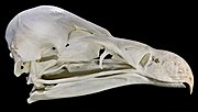 Vulture skull
 -
 King cobra skull
 -
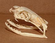
 Goat skull
 -
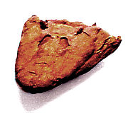 Skull oflobe-finned fishand early tetrapods
 -
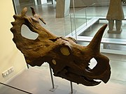 Centrosaurus skull
.jpg)


See also
- Craniometry
- Crystal skull
- Head and neck anatomy
- Human skull symbolism
- Memento mori
- Plagiocephaly, the abnormal flattening of one side of the skull
- Skull and crossbones (disambiguation)
- Teshik-Tash
- Totenkopf
- Yorick
- Overmodelled skull
- Diploë
References
![]() This article incorporates text in the public domain from page 128 of the 20th edition of Gray's Anatomy (1918)
This article incorporates text in the public domain from page 128 of the 20th edition of Gray's Anatomy (1918)
- Merriam-Webster Dictionary. Archivedfrom the original on 17 February 2015.
- ISBN 9780080920856.
- ^ "Cephalization: Biology". Encyclopædia Britannica. Archived from the original on 2 May 2016. Retrieved 23 April 2016.
- ^ "Definition of skull | Dictionary.com". www.dictionary.com. Retrieved 6 September 2021.
- ISBN 9780375763427.
- ISBN 9783030153632.
- ^ "Jugal Bone – an overview | ScienceDirect Topics".
- ^ ISBN 0-03-910284-X.
- ^ ISBN 0-03-910284-X.
- ^ Wing, Leonard W. (1956). "The Place of Birds in Nature". Natural History of Birds. The Ronald Press Company. pp. 22–23.
- ISBN 0-8151-1458-3.
- PMID 14507064. Archived from the originalon 24 September 2011.
- ^ ISBN 978-81-239-2332-1.
- ^ Silva, Sandra; Jeanty, Philippe (7 June 1999). "Cloverleaf skull or kleeblattschadel". TheFetus.net. MacroMedia. Archived from the original on 13 February 2008. Retrieved 3 February 2007.
- ^ S2CID 34344899.
- PMID 1513883.
- ^ Gaillard, Frank. "Copper beaten skull". Radiopaedia. Archived from the original on 25 April 2018. Retrieved 25 April 2018.
- .
- ^ "Repeated Concussions Can Thicken the Skull". 2 September 2022.
- ^ "3D-Printed Polymer Skull Implant Used For First Time in US". Medical Daily. 7 March 2013. Archived from the original on 28 September 2013. Retrieved 24 September 2013.
- ^ "Dutch hospital gives patient new plastic skull, made by 3D printer". DutchNews.nl. 26 March 2014. Archived from the original on 28 March 2014.
- ^ Cohut, Maria (29 August 2018). "Newly discovered skull channels play role in immunity". Medical News Today. Retrieved 30 August 2018.
- S2CID 601504.
- PMID 19198644.
- ^ World Professional Association for Transgender Health. WPATH Clarification on Medical Necessity of Treatment, Sex Reassignment, and Insurance Coverage in the U.S.A. Archived 30 September 2011 at the Wayback Machine (2008).
- ^ World Professional Association for Transgender Health. Standards of Care for the Health of Transsexual, Transgender, and Gender Nonconforming People, Version 7. Archived 3 March 2012 at the Wayback Machine pg. 58 (2011).
- ^ PMID 394780.
- ^ "5d. The Interior of the Skull". Gray's Anatomy. Archived from the original on 31 March 2014. Retrieved 22 October 2014.
- ^ Other Sources:
- Li, Haiyan; Ruan, Jesse; Xie, Zhonghua; Wang, Hao; Liu, Wengling (2007). "Investigation of the critical geometric characteristics of living human skulls utilising medical image analysis techniques". International Journal of Vehicle Safety. 2 (4): 345. .
- name="Men May Be More Susceptible To Head Injury Than Women, Study Suggests">"Men May Be More Susceptible To Head Injury Than Women, Study Suggests". ScienceDaily. 22 January 2008. Archived from the original on 7 March 2012. Retrieved 6 June 2012.
- De Boer, H. H. (Hans); Van der Merwe, A. E. (Lida); Soerdjbalie-Maikoe, V. (Vidija) (September 2016). "Human cranial vault thickness in a contemporary sample of 1097 autopsy cases: relation to body weight, stature, age, sex and ancestry". International Journal of Legal Medicine. 130 (5): 1371–1377. PMID 26914798.
- Ross, M. D.; Lee, K. A.; Castle, W. M. (10 April 1976). "Skull thickness of Black and White races". South African Medical Journal = Suid-Afrikaanse Tydskrif vir Geneeskunde. 50 (16): 635–638. PMID 1224277.
- Adeloye, Adelola; Kattan, Kenneth R.; Silverman, Frederic N. (July 1975). "Thickness of the normal skull in the American blacks and whites". American Journal of Physical Anthropology. 43 (1): 23–30. PMID 1155589.
- "International Journal of Research in Medical Sciences". www.msjonline.org. Retrieved 18 February 2021.
- Ekşi, Murat Şakir; Güdük, Mustafa; Usseli, Murat Imre (19 November 2020). "Frontal Bone is Thicker in Women and Frontal Sinus is Larger in Men: A Morphometric Analysis". The Journal of Craniofacial Surgery. 32 (5): 1683–1684. S2CID 227159148.
- PMID 16364185.
- S2CID 33825332.
- PMID 26255873.
- S2CID 39294670.
- PMID 24551607.
- OCLC 50485765.
- OCLC 43662032.
External links
- Skull Module (California State University Department of Anthology)
- Skull Anatomy Tutorial. (GateWay Community College)
- Bird Skull Collection Bird skull database with very large collection of skulls (Agricultural University of Wageningen)
- Human skull base (in German)
- Human Skulls / Anthropological Skulls / Comparison of Skulls of Vertebrates (PDF; 502 kB)