
Sponge spicule
similar spicules
Spicules are structural elements found in most
Sponge spicules are made of
Overview
| Part of a series related to |
| Biomineralization |
|---|
 |
Sponges are a species-rich clade of the earliest-diverging (most basal) animals.[3] They are distributed globally,[4] with diverse ecologies and functions,[5][6][7][8][9][10] and a record spanning at least the entire Phanerozoic.[11]
Most sponges produce skeletons formed by spicules, structural elements that develop in a wide variety of sizes and three dimensional shapes. Among the four sub-clades of Porifera, three (
Research history
In 1833,
Later, the
Then, during the German Deep Sea Expedition "
Since their discovery, hexactinellids were appraised as "the most characteristic inhabitants of the great depths", rivalling in beauty the other class of siliceous Porifera, the demosponges.[20] Their thin network of living tissues is supported by a characteristic skeleton, a delicate scaffold of siliceous spicules, some of which may be fused together by secondary silica deposition to form a rigid framework.[21] The Hexactinellida together with the Demospongiae forms a common taxonomic unit comprising the siliceous sponges. The spicules, the elements from which their skeletons are constructed, are built in a variety of distinct shapes, and are made from silica that is deposited in the form of amorphous opal (SiO2·nH2O).[19]
In evolution, after the Ediacaran period, a third class of Porifera appeared, the
Sponges have been receiving special attention from researchers since the introduction of
Spicule types
Sponge spicules can be calcareous or siliceous. Siliceous spicules are sometimes embedded in spongin. Spicules are found in a range of symmetry types.


Monaxons form simple
Triaxons have three axes; in triods, each axis bears a similar ray; in pentacts, the triaxon has five rays, four of which lie in a single plane; and pinnules are pentacts with large spines on the non-planar ray.[31]
Tetraxons have four axes, and polyaxons more (description of types to be incorporated from [31]). Sigma-C spicules have the shape of a C.[31]
Dendroclones might be unique to extinct sponges[32] and are branching spicules that may take irregular forms, or may form structures with an I, Y or X shape.[33][34]
- Megascleres are large spicules measuring from 60-2000 μm and often function as the main support elements in the skeleton.[35]
- Acanthostyles are spiny styles.
- Anatriaenes, orthotriaenes and protriaenes are triaenes[36] - megascleres with one long and three short rays.
- Strongyles are megascleres with both ends blunt or rounded.
- Styles are megascleres with one end pointed and the other end rounded.
- Tornotes are megascleres with spear shaped ends.
- Tylotes are megascleres with knobs on both ends.
- Microscleres are small spicules measuring from 10-60 μm and are scattered throughout the tissue and are not part of the main support element.[35]
- Chelae are microscleres with shovel-like structures on the ends. Anisochelas are microscleres with dissimilar ends. Isochelas are microscleres with two similar ends.
- Euasters are star-shaped microscleres with multiple rays radiating from a common centre. Examples are oxyasters (euasters with pointed rays) or sterrasters (ball-shaped euasters).
- Forceps are microscleres bent back on themselves.
- Microstrongyles are small rods with both ends blunt or rounded.
- Microxeas are small rods with both ends pointed.
- Sigmas are C- or S-shaped microscleres.
Calcareous spicules
Animal biomineralization is a controlled process and leads to the production of mineral–organic composite materials that considerably differ in shape and material properties from their purely inorganic counterparts. The ability to form functional biominerals, such as endoskeletons and exoskeletons, protective shells, or teeth, had been a significant step in animal evolution. Calcium carbonate biomineralization, the most widespread type among animal phyla,[37] evolved several times independently, resulting in multiple recruitments of the same genes for biomineralization in different lineages.[38]
Among these genes, members of the
Among extant sponges, only the

Spicules are formed by sclerocytes, which are derived from archaeocytes. The sclerocyte begins with an organic filament, and adds silica to it. Spicules are generally elongated at a rate of 1-10 μm per hour. Once the spicule reaches a certain length it protrudes from the sclerocyte cell body, but remains within the cell's membrane. On occasion, sclerocytes may begin a second spicule while the first is still in progress.[51]
The shapes of calcareous sponge spicules are simple compared with the sometimes very elaborate siliceous spicules found in the other sponge classes. With only a few exceptions, calcareous sponge spicules can be of three basic types: monaxonic, two-tipped diactines, triactines with three spicules rays, and four-rayed tetractines. Specialized cells, the sclerocytes, produce these spicules, and only a few sclerocytes interact in the formation of one specific spicule: Two sclerocytes produce a diactine, six sclerocytes form a triactine, and seven a tetractines.[52][53][54] A pair of sclerocytes is involved in the growth of each actine of these spicules. After an initial phase, the so-called founder cell promotes actine elongation, the second, so-called thickener cell in some, but not all species deposit additional calcium carbonate on the actine, as it migrates back toward the founder cell.[54][55] Calcareous sponges can possess only one or any combination of the three spicule types in their body, and in many cases, certain spicule types are restricted to specific body parts. This indicates that spicule formation is under strict genetic control in calcareous sponges, and specific CAs play an essential role in this genetic control[50][48]
Siliceous spicules
The largest biosilica structure on Earth is the giant basal spicule from the deep-sea
- (a) Young specimens of M. chuni anchored to the muddy substratum by one single giant basal spicule (gbs). The body (bo) surrounds the spicule as a continuous, round cylinder.
- (b) The growth phases of the sessile animal with its GBS (gbs) which anchors it to the substratum and holds the surrounding soft body (bo). The characteristic habitus displays linearly arranged large atrial openings (at) of approximately 2 cm in diameter. With growth, the soft body dies off in the basal region and exposes the bare GBS (a to c).
- (c) Part of the body (bo) with its atrial openings (at). The body surface is interspersed with ingestion openings allowing a continuous water flow though canals in the interior which open into oscules that are centralized in atrial openings, the sieve-plates.
- (d) M. chuni in its natural soft bottom habitat of bathyal slopes off New Caledonia. The specimens live at a depth of 800–1,000 m [23]. In this region, the sponge occurs at a population density of 1-2 individuals per m2. The animals reach sizes of around 1 m in length.
- (e) Drawings of different glass sponges (hexactinellids).[19]

Not to scale — sizes vary between 0.01 and 1 mm

Geodia atlantica; (h) is the hilum; the arrow points to a smooth rosette made up of rays. Left scale 20 μm, right scale 2 μm.

Geodia macandrewii, left scale 50 μm, right scale 2 μm
Siliceous spicules in demosponges exist in a variety of shapes, some of which look like minute spheres of glass. They are called sterrasters when they belong to the Geodiidae family and selenasters when they belong to the Placospongiidae family.[58]
Siliceous spicules were first described and illustrated in 1753 by
Today, the Geodiidae represent a highly diverse sponge family with more than 340 species, occurring in shallow to deep waters worldwide apart from the Antarctic. Sterrasters/aspidaster spicules are currently the main
Selenasters are the main
Sterrasters/selenasters are big enough to examine in some detail their surfaces with an optical microscope. However, the use of the scanning electron microscope (SEM) enabled a significantly better understanding of the surface microornamentations. A few descriptive terms have also appeared to describe and compare in greater detail the microornamentations of these ball-shaped spicules. polyaxial spicules such as the sterrasters and aspidasters, are the result of fused "actines" (= branches of asters, from the Greek for "star"), later covered with "rosettes" made of different "rays". The "hilum" (Latin for a "little thing" or "trifle" or the "eye of a bean") is a small area without rosettes or any kind of surface pattern. There are no particular terms to describe the surface of selenasters, except for the "hilum", also present. Although there appears to be no significant variation in the size of the rosettes and hilum between species,[70][67] noticed that rosettes could be smooth or warty and hypothesized that this character could be of phylogenetic value if studied more broadly. Furthermore, the rosette morphology also seemed to be variable between Geodia, Pachymatisma, and Caminella [71][72] which suggests that a more detailed study of the sterraster/aspidaster surface would potentially bring new characters for Geodiidae genera identification.[58]
Spicule "life cycle"

From formation to deposition
The formation of spicules is controlled genetically.[73] In most cases, the first growth phase is intracellular; it starts in sclerocytes (amoeboid cells responsible for spicule formation) in mesohyl [74][75] and is mediated by silicatein, a special enzyme that initiates formation of the axial filament (harboured by the axial canal) which provides the vertical axis of the spicule.[76] The axial canal is filled with organic proteinaceous material which usually extends to the tip of the newly-formed spicule.[77] The cross-section of the axial canal differs across major sponge clades that produce siliceous spicules (it is triangular in demosponges,[78] irregular in homoscleromorphs [14] and quadrangular in hexactinellids.[79] In calcareans (producing calcareous spicules) the axial canal is not developed.[79] The geometry and the length of the axial filament determines the shape of the spicule.[80] In desmoid spicules of 'lithistids' (an informal group of demosponges with articulated skeletons), however, the axial filament is shorter than the spicule arms and it is possible that only organic molecules are involved in the spicule-forming process.[80][16]
During formation of the siliceous spicules (Calcarea displays different mechanisms of spicule biomineralization), sponges obtain silicon in the form of soluble silicic acid and deposit it around the axial filament, [78][14] within a special membrane called silicalemma.[81][82] Silica is first laid out as small 2 μm granules [78][80] that are fused to bigger spheres (or fused together within process of biosintering in Hexactinellida.[83] After some time, amorphous silica is added, forming evenly-deposited concentric layers,[14] separated from each other by ultrathin organic interlayers.[84] At this stage, immature spicules are secreted from the sclerocyte and covered by pseudopodia of one to several cells, and the process of silica deposition and spicule growth continues.[78][16]
After completing the deposition of silica (or during this phase), the spicule is transported to the right place in the sponge body by crawling mesohyl cells, where spongocytes secrete spongin fibrils around them and connect them with adjacent spicules.[14] In some hexactinellids, that are characterized by rigid skeleton, the fusion of spicules appears to occur parallel to spicule secretion.[85][16]
When sponges are alive, their spicules provide a structural "framework". Following their death, the body and the skeleton structure, especially that of demosponges in which the spicules are connected to each other only by perishable collagen fibres, rapidly disintegrate leaving the spicules "free". Because of this, sponges are rarely wholly preserved in the fossil record. Their spicules, however, are incorporated into sediments, often becoming one of the main components of sedimentary rocks.[86][87] Sometimes spicules accumulate into enormous agglomerations called spicule mats or beds.[88] These accumulations are characteristic for polar waters.[89][90] Spicules can fossilize to form special type of rocks called the spiculites ("spongillites" for freshwater sponge spicules); these types of rocks are known globally,[91][92][93][94] and have been formed through the whole Phanerozoic.[92] Biosiliceous sedimentation occasionally results in the formation of spiculitic cherts (in so called glass ramps) which are recorded from the Permian to Eocene of many parts of the world.[95][96][16]
Locomotion
In 2016 a newly discovered demosponge community living under arctic ice were found to have moved across the sea floor by extending their spicules and then retracting their body in the direction of motion.[97]
Spiculites
When dead sponge bodies disintegrate, spicules become incorporated into
The record of fossil and subfossil sponge spicules is extraordinarily rich and often serves as a basis for far-reaching reconstructions of sponge communities, though spicules are also bearers of significant ecological and environmental information. Specific requirements and preferences of sponges can be used to interpret the environment in which they lived, and reconstruct oscillations in water depths, pH, temperatures, and other parameters, providing snapshots of past climate conditions. In turn, the
Spicules provide structural support for maintaining the vertical body position, minimize the metabolic cost of water exchange,
The mineral composition of sponge spicules makes these structures the most resistant parts of the sponge bodies [79] and ensures the ability of spicules to withstand various taphonomic processes,[86][101] resulting in that they often constitute the only evidence of the presence of some sponges in an ecosystem.[102] Even though sponges are often known from rich assemblages of bodily-preserved specimens,[103][104][105] a significant part of their fossil and subfossil record is also represented by their spicules. Having that in mind, spicules can be of crucial importance for reconstructions of extinct or cryptic (hiding in cervices and caves) sponge communities; and, indeed, they have been investigated especially with respect to their taxonomic significance.[106][12] The morphologies of spicules and their arrangement, together with other important sponge features, such as the shape, consistency, and color, are essential when identifying sponges.[107][16]
In contrast to whole-bodied sponge fossils, spicules are common in many depositional environments.[108] Their significance, however, is often underestimated, which is mostly due to the difficulties in assigning disassociated spicules to sponge taxa or due to the scarcity of the material.[16]
Interaction with light
Research on the
-
 Sponge spicules
 -
 Spicules of sponge (SEM)
 -
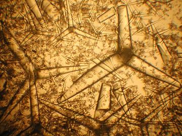
 Network of sponge spicules

.JPG)

See also
References
- S2CID 85614908.
- .
- PMID 22560777.
- PMID 22558119.
- S2CID 41590456.
- .
- .
- PMID 22664122.
- S2CID 6720678.
- ISBN 978-3-319-59007-3.
- ^ ISBN 978-0-306-47260-2.
- ^ ISBN 978-0-306-47260-2.
- PMID 24487057.
- ^ S2CID 535640.
- PMID 17360189.
- ^ PMID 33384908..
 Material was copied from this source, which is available under a Creative Commons Attribution 4.0 International License
 - ^ a b Grant, R. E. (1833). "Lectures on comparative anatomy and animal physiology; on the classification of the animals". The Lancet. 1: 153–159.
- ISBN 9780785503712.
- ^ PMID 21941585.. Material was copied from this source, which is available under a Creative Commons Attribution 4.0 International License
- ISBN 9781378320709.
- ^ PMID 17298890.
- PMID 10081159.
- PMID 9402742.
- S2CID 10557258.
- S2CID 27535038.
- S2CID 29453726.
- S2CID 28844289.
- doi:10.2113/0540329.
- ISBN 9783527600908.)
{{cite journal}}: CS1 maint: DOI inactive as of January 2024 (link - ^ Pier, J. Q. (2019) "Porifera" Digital Atlas of Ancient Life, Paleontological Research Institution, New York. Updated: 16 Sept 2020.
- ^ a b c d Eugenio Andri; Stefania Gerbaudo; Massimiliano Testa (2001). "13. Quaternary siliceous sponge spicules in the western Woodlark basin, southwest Pacific (ODP LEG 180)" (PDF). In Huchon, P.; Taylor, B.; Klaus, A (eds.). Proceedings of the Ocean Drilling Program, Scientific Results. Vol. 180. pp. 1–8.
- ^ Komal Sutar and Bhavika Bhoir. "Study of the sponge spicules from the coastline of Raigad district, Maharashtra, India" (PDF). vpmthane.org/.
- S2CID 130654425.
- S2CID 130617208.
- ^ .
- ^ Boury-Esnault, Nicole, and Klaus Rutzler, editors. Thesaurus of Sponge Morphology. Smithsonian Contributions to Zoology, number 596, 55 pages, 305 figures, 1997. https://www.portol.org/thesaurus
- S2CID 45466684.
- S2CID 218872873.
- ^ S2CID 4846408.
- PMID 11696553.
- PMID 24146378.
- PMID 32917908.
- PMID 27407171.
- PMID 22607884.
- ^ PMID 25421146.
- S2CID 7042860.
- PMID 26536128.
- ^ PMID 33897759.. Material was copied from this source, which is available under a Creative Commons Attribution 4.0 International License
- doi:10.1139/z06-005.
- ^ PMID 28406140.
- ^ S2CID 22067910.
- .
- .
- ^ S2CID 9916021.
- S2CID 85345222.
- ISSN 1618-3193. Archived from the original(PDF) on 2011-07-24. Retrieved 2009-05-25.
- PMID 22558119.
- ^ doi:10.3389/fmars.2020.613610.. Material was copied from this source, which is available under a Creative Commons Attribution 4.0 International License
- ^ Donati, V. (1753). "Auszug seiner Natur-Geschichte des Adriatischen Meeres", Halle: Palala Press.
- ^ a b Sollas, W. J. (1888). "Report on the Tetractinellida collected by H.M.S. Challenger, during the years 1873-1876". Report on the Scientific Results of the Voyage of H.M.S. Challenger, 1873-1876. Zoology, 25: 1–458.
- ^ Hanitsch, R. (1895). "Notes on a collection of sponges from the West Coast of Portugal". Trans. Liverpool Biol. Soc., 9: 205–219.
- ^ von Lendenfeld, R. (1910). "The Sponges. 2. The erylidae". In: Reports on the scientific results of the expedition to the eastern tropical pacific, in charge of Alexander Agassiz, by the U.S. Fish Commission Steamer 'Albatross', from October, 1904, to March, 1905, Lieut. Commander L.M. Garrett, U.S.N., Commanding, and of other Expeditions of the Albatross, 1888-1904. (21). Mem. Museum Comp. Zool., 41: 261–324.
- S2CID 85137461.
- PMID 21494664.
- ^ Santucci, R. (1922). "La Geodia cydonium come centro di associazione biologica". Mem. Comitato Talassografico ltaliano: 103: 5–19.
- ^ Corriero, G. (1987) (1989). "The sponge fauna from the Stagnone di Marsala (Sicily): taxonomic and ecological observations". Bollettino dei musei e degli istituti biologici dell, Univ. Genova, 53: 101–113.
- ^ S2CID 85922551.
- ^ PMID 23794884.
- ^ Hajdu, E., Peixinho, S., and Fernandez, J. C. C. (2011). "Esponjas Marinhas da Bahia - Guia de Campo e Laboratório". Série Livros, 45. Rio de Janeiro: Museu Nacional/UFRJ.
- .
- .
- S2CID 52976107.
- PMID 10903523.
- S2CID 39893025.
- S2CID 12720013.
- PMID 9600948.
- S2CID 35458775.
- ^ S2CID 21428171.
- ^ doi:10.1139/z06-032.
- ^ S2CID 26495972.
- ISBN 9781461252146.
- ISBN 978-1-4757-6116-0.
- PMID 19683578.
- PMID 26094876.
- ISBN 978-0-306-47260-2.
- ^ S2CID 86814441.
- JSTOR 3226383.
- .
- ^ Koltun, V.M. (1968). "Spicules of sponges as an element of the bottom sediments of the Antarctic". In: SCAR Symposium on Antarctic Oceanography, Scott Polar Research Institute, pages 21–123.
- S2CID 129803464.
- ^ Cayeux L. (1929). "Les roches sédimentaires de France: roches siliceuses". Mémoires de la Carte Géologique de France 1929, pages 1-774
- ^ a b Gammon, P.R., Middleton, G.V., Church, M.J., Coniglio, M., Hardie, L.A. and Longstaffe, F.J. (1978) "Spiculites and spongolites}. In: Encyclopedia of Sediments and Sedimentary Rocks, Dordrecht: Springer.
- .
- S2CID 129770411.
- .
- S2CID 134003011.
- S2CID 233429396.
- S2CID 28635129.
- ISBN 978-0-306-47260-2.
- ISBN 978-0-306-47260-2.
- S2CID 2072201.
- S2CID 9536558.
- ISBN 978-3-642-75658-0.
- .
- S2CID 89387705.
- ^ Diaz, M.C. and Rützler, K. (2001). "Sponges: an essential component of Caribbean coral reefs". Bulletin of marine science, 69(2): 535–546.
- ^ Collin, R., Diaz, M.C., Norenburg, J.L., Rocha, R.D., Sanchez, J.A., Schulz, A., Schwartz, M.L. and Valdes, A. (2005) "Photographic identification guide to some common marine invertebrates of Bocas Del Toro, Panama". Caribbean Journal of Science.
- doi:10.1139/z05-169.
- PMID 14993612.
- PMID 14993612.
- .
- ^ "Nature's 'fibre optics' experts". BBC. 2008-11-10. Retrieved 2008-11-10.
Further references
- Shoham, Erez; Prohaska, Thomas; Barkay, Zahava; Zitek, Andreas; Benayahu, Yehuda (2019). "Soft corals form aragonite-precipitated columnar spiculite in mesophotic reefs". Scientific Reports. 9 (1): 1241. PMID 30718658.