
Spike protein
In virology, a spike protein or peplomer protein is a protein that forms a large structure known as a spike or peplomer projecting from the surface of an enveloped virus.[2][3]: 29–33 The proteins are usually glycoproteins that form dimers or trimers.[3]: 29–33 [4]
History and etymology
The term "peplomer" refers to an individual spike from the viral surface; collectively the layer of material at the outer surface of the
Properties
Spikes or peplomers are usually rod- or club-shaped projections from the viral surface. Spike proteins are
Functions
Spikes typically have a role in
Examples
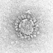
Spikes or peplomers can be visible in
The COVID-19 pandemic necessitated identification of viral particles in electron micrographs of patient tissue samples. A number of reports misidentified normal subcellular structures as coronaviruses due to their superficial resemblance to coronavirus morphology, and because the distinctive spikes of coronaviruses are apparent by negative stain but much less visible in thin section.[14]
Influenza viruses
Most
Retroviruses
Gallery
-
-
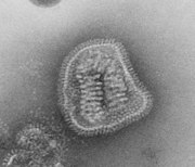 Influenza virus
 -
 Human immunodeficiency virus


See also
References
- ^ Solodovnikov, Alexey; Arkhipova, Valeria (29 July 2021). "Достоверно красиво: как мы сделали 3D-модель SARS-CoV-2" [Truly beautiful: how we made the SARS-CoV-2 3D model] (in Russian). N+1. Archived from the original on 30 July 2021. Retrieved 30 July 2021.
- The Free Dictionary. Farlex. 2011. Retrieved 30 March 2011.
- ^ ISBN 978-0123751560.)
{{cite book}}: CS1 maint: location missing publisher (link - ISBN 9780128145166.
- ^ PMID 5330240.
- ^ ISBN 9780080920368.
- PMID 14467544.
- PMID 13931895.
- PMID 25866377.
- PMID 32833200.
- S2CID 221503034.
- PMID 33619260.
- PMID 21994708.
- PMID 33600302.
- PMID 22864288.