
Submucosa
This article needs additional citations for verification. (March 2013) |
| Submucosa | |
|---|---|
longitudinal muscle Serosa or Adventitia | |
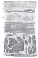
 Endoscopy and radial endoscopic ultrasound images of submucosal tumour in mid-esophagus. The submucosa is seen as a dark ring on the ultrasound image. | |
| Details | |
| Identifiers | |
| Latin | tela submucosa |
| TA98 | A05.3.01.028 A05.4.01.014 A05.5.01.026 A05.6.01.008 A05.7.01.005 A06.4.02.028 A08.3.01.022 |
| TA2 | 2857, 2891, 2913, 2939, 2967, 3221, 3234, 3419 |
| FMA | 85391 |
| Anatomical terminology] | |
The submucosa (or tela submucosa) is a thin layer of
The submucosa (sub- + mucosa) is to a mucous membrane what the subserosa (sub- + serosa) is to a serous membrane.
Structure
In the
Clinical significance
Identification of the submucosa plays an important role in diagnostic and therapeutic
The submucosa is also identified in
Endoscopic mucosal resection involves removal of the mucosal layer, and in order to be done safely, a submucosal injection of dye is performed to ensure integrity at the beginning of the procedure.
Female uterine submucosal layers are liable to develop fibroids during pregnancy and are often excised upon discovery.[1]
Small intestinal submucosa
Small intestinal submucosa (SIS) is submucosal
Unlike other scaffold materials, the resorbable SIS extracellular matrix (SIS-ECM) scaffold is replaced by well-organized host tissues, including differentiated skeletal muscle.[4]
History
A scientific article published in March 2018[5] proposed a revision of the anatomical definition of the submucosa. They first saw a non compact tissue which should be submucosa using a technology called endomicroscopy. They hypothesised that the submucosa was not compact as it was previously seen on histological analysis but form a reticular pattern. To confirm their findings, they performed fixed samples of bile duct into a freezing media in order to conserve the shape of the submucosa. They then performed a histological analysis and with several staining techniques, they described the submucosa as a network of collagenous bands separating open, formerly fluid-filled spaces. Theses spaces are bordered by fibroblast-like cells CD34 positive. However, these cells are devoid of ultrastructural features indicative of endothelial differentiation, including pinocytotic vesicles and Weibel-Palade bodies.
Additional images
-
 Stomach.
 -
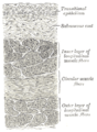
 Section of the human esophagus. Moderately magnified.
 -
 Vertical section of bladder wall.
 -
 General structure of the gut wall showing the submucosa.


References
- ^ "What is the submucosa and why is it important? - Submucosa". Archived from the original on 2019-06-23. Retrieved 2018-07-25.
- PMID 16998356.
- PMID 15967902.
- PMID 11922734.
- ^ Benias, P., Wells, R., Sackey-Aboagye, B., Klavan, H., Reidy, J., Buonocore, D., Miranda, M., Kornacki, S., Wayne, M., Carr-Locke, D. and Theise, N. (2018). Structure and Distribution of an Unrecognized Interstitium in Human Tissues. Scientific Reports, 8(1).