
Superior laryngeal nerve
| Superior laryngeal nerve | |
|---|---|
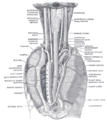
 Plan of upper portions of glossopharyngeal nerve, vagus nerve, and accessory nerve. ("Laryngeal" labeled at lower right.) | |
 Course and distribution of the glossopharyngeal, vagus, and accessory nerves. (Branches visible in upper right.) | |
| Details | |
| From | Vagus nerve |
| Innervates | larynx |
| Identifiers | |
| Latin | nervus laryngeus superior |
| TA98 | A14.2.01.160 |
| TA2 | 6339 |
| FMA | 6239 |
| Anatomical terms of neuroanatomy | |
The superior laryngeal nerve is a branch of the vagus nerve. It arises from the middle of the inferior ganglion of vagus nerve and additionally also receives a sympathetic branch from the superior cervical ganglion.
The superior laryngeal nerve produces of two branches: the internal laryngeal nerve (its sensory branch) which supplies sensory fibers to the laryngeal mucosa, and the external laryngeal nerve (its motor branch) which innervates the cricothyroid muscle.
Structure
Origin
The superior laryngeal nerve arises from the middle of the inferior ganglion of vagus nerve.
Course
The superior laryngeal nerve descends by the side of the pharynx deep to the internal carotid artery before dividing into two branches —the external laryngeal nerve and the internal laryngeal nerve.[1]
Branches
External laryngeal nerve
The external laryngeal nerve is the smaller, external branch. It descends on the
Internal laryngeal nerve
The internal laryngeal nerve is the internal branch. It descends to the thyrohyoid membrane, piercing it in company with the superior laryngeal artery, and is distributed to the mucous membrane of the larynx. Of these sensory branches, some are distributed to the epiglottis, the base of the tongue, and the epiglottic glands; others pass posteriorly, in the aryepiglottic fold, to supply the mucous membrane surrounding the entrance of the larynx, and the mucous lining of the larynx as far down as the vocal folds.
A filament descends beneath the mucous membrane on the inner surface of the thyroid cartilage and joins the recurrent laryngeal nerve. Above the vocal folds the sensory innervation of the larynx is via the internal laryngeal nerve. Below the vocal folds it is by way of branches of the recurrent laryngeal nerve. The vocal fold itself receives dual innervation from both nerves.
Function
The superior laryngeal nerve innervates the cricothyroid muscle.[2]
Clinical significance
A superior laryngeal nerve palsy changes the pitch of the voice and causes an inability to make explosive sounds due to paralysis of the cricothyroid muscle. If no recovery is evident three months after the palsy initially presents, the damage is most likely to be permanent. A bilateral palsy presents as a tiring and hoarse voice. It can be injured in surgery involving the removal of the
Irritation of the internal laryngeal nerve results in uncontrolled coughing - usually as a result of food or water in the
Additional images
-
 The position and relation of the esophagus in the cervical region and in the posterior mediastinum. Seen from behind.

References
![]() This article incorporates text in the public domain from page 912 of the 20th edition of Gray's Anatomy (1918)
This article incorporates text in the public domain from page 912 of the 20th edition of Gray's Anatomy (1918)
- ISBN 978-0-7295-3752-0.
- ISBN 978-0-323-04019-8, retrieved 2021-01-12
- S2CID 8105192.
- S2CID 29270084.
- PMID 11886347.
- PMID 29142848.
- PMID 16402208.
External links
- cranialnerves at The Anatomy Lesson by Wesley Norman (Georgetown University) (X)