
Superior rectal artery
| Superior rectal artery | |
|---|---|
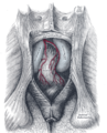
anastomoses. (Superior hemorrhoidal artery visible at center right.) | |
 | |
| Details | |
| Source | Inferior mesenteric artery |
| Vein | Superior rectal vein |
| Supplies | Rectum |
| Identifiers | |
| Latin | arteria rectalis superior, arteria haemorrhoidalis superior |
| TA98 | A12.2.12.073 |
| TA2 | 4296 |
| FMA | 14832 |
| Anatomical terminology] | |
The superior rectal artery (superior hemorrhoidal artery) is an artery that descends into the pelvis to supply blood to the rectum.
Structure
The superior rectal artery is the continuation of the inferior mesenteric artery. It descends into the pelvis between the layers of the mesentery of the sigmoid colon, crossing the left common iliac artery and vein.
It divides, opposite the third
These pierce the muscular coat of the bowel and run downward, as straight vessels, placed at regular intervals from each other in the wall of the gut between its muscular and mucous coats, to the level of the internal anal sphincter; here they form a series of loops around the lower end of the rectum, and communicate with the middle rectal artery (from the internal iliac artery) and with the inferior rectal artery (from the internal pudendal artery).
Function
The superior rectal artery supplies the rectum and the anus.[1][2]
Additional images
-
 The posterior aspect of the rectum exposed by removing the lower part of the sacrum and the coccyx.

See also
References
![]() This article incorporates text in the public domain from page 610 of the 20th edition of Gray's Anatomy (1918)
This article incorporates text in the public domain from page 610 of the 20th edition of Gray's Anatomy (1918)
- ^ ISBN 978-0-7020-3367-4, retrieved 2021-02-03
- ISBN 978-0-443-10373-5, retrieved 2021-02-03
External links
- Anatomy figure: 39:02-06 at Human Anatomy Online, SUNY Downstate Medical Center - "Branches of the inferior mesenteric artery."
- Anatomy photo:39:05-0112 at the SUNY Downstate Medical Center - "Intestines and Pancreas: Branches of the Inferior Mesenteric Artery"
- sup&infmesentericart at The Anatomy Lesson by Wesley Norman (Georgetown University)
- pelvis at The Anatomy Lesson by Wesley Norman (Georgetown University) (pelvicarteries)