
Taenia solium
| Taenia solium | |
|---|---|

| |
| Scolex (head) of Taenia solium | |
| Scientific classification | |
| Domain: | Eukaryota |
| Kingdom: | Animalia |
| Phylum: | Platyhelminthes |
| Class: | Cestoda |
| Order: | Cyclophyllidea |
| Family: | Taeniidae |
| Genus: | Taenia |
| Species: | T. solium
|
| Binomial name | |
| Taenia solium | |
Taenia solium, the pork tapeworm, belongs to the
There are two forms of human infection. One is "primary hosting", called taeniasis, and is due to eating under-cooked pork that contains the cysts and results in adult worms in the intestines. This form generally is without symptoms; the infected person does not know they have tapeworms. This form is easily treated with anthelmintic medications which eliminate the tapeworm. The other form, "secondary hosting", called cysticercosis, is due to eating food, or drinking water, contaminated with faeces from someone infected by the adult worms, thus ingesting the tapeworm eggs, instead of the cysts. The eggs go on to develop cysts primarily in the muscles, and usually with no symptoms. However some people have obvious symptoms, the most harmful and chronic form of which is when the cysts form in the brain. Treatment of this form is more difficult but possible.
The adult worm has a flat, ribbon-like body which is white and measures 2 to 3 metres (6' to 10') long, or more. Its tiny attachment, the
Human primary hosting is best diagnosed by microscopy of eggs in faeces, often triggered by spotting shed segments. In secondary hosting, imaging techniques such as
T. solium deeply affects developing countries, especially in rural settings where pigs roam free,[1] as clinical manifestations are highly dependent on the number, size, and location of the parasites as well as the host's immune and inflammatory response.[2]
Description
Adult T. solium is a
After a short neck is the elongated body, the strobila. The entire body is covered by a covering called a
If released early enough in the digestive tract and not passed, fertilised eggs can mature using upper tract digestive enzymes and the tiny larvae migrate to form
-
 Taenia solium adult
 -
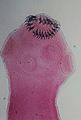 Taenia solium scolex (x400)
 -
 Egg of T. solium


Life cycle
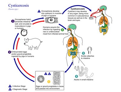
The life cycle of T. solium is indirect as it passes through pigs, as intermediate hosts, into humans, as definitive hosts. In humans the infection can be relatively short or long lasting, and in the latter case if reaching the brain can last for life. From humans, the eggs are released in the environment where they await ingestion by another host. In the secondary host, the eggs develop into oncospheres which bore through the intestinal wall and migrate to other parts of the body where the cysticerci form. The cysticerci can survive for several years in the animal.[15]
Definitive host
Humans are colonised by the larval stage, the cysticercus, from undercooked pork or other meat. Each microscopic cysticercus is oval in shape, containing an inverted scolex (specifically "protoscolex"), which everts once the organism is inside the small intestine. This process of
As a hermaphrodite, it reproduces by
Intermediate host
Pigs are the principal intermediate hosts that ingest the eggs in traces of human faeces, mainly from vegetation contaminated with it such as from water bearing traces of it. The embryonated eggs enter intestine where they
Humans are also accidental secondary hosts when they are colonised by embryonated eggs, either by auto-colonisation or ingestion of contaminated food. As in pigs, the oncospheres hatch and enter blood circulation. When they settle to form cysts, clinical symptoms of cysticercosis appear. The cysticercus is often called the metacestode.[21]
Diseases
Signs and symptoms
Taeniasis
Taeniasis is infection in the intestines by the adult T. solium. It generally has mild or
These symptoms could continue until the tapeworm dies from the course of treatment but otherwise could continue for many years, as long as the worm lives. If untreated it is common that the infections with T. solium last for approximately 2–3 years. It is possible that infected people may show no symptoms for years.[22]
Cysticercosis
Ingestion of T. solium eggs or egg-containing proglottids which rupture within the host intestines results in the development and subsequent migration of larvae into host tissue to cause cysticercosis. In pigs, there are not normally pathological lesions as they easily develop immunity.[23] But in humans, infection with the eggs causes serious medical conditions. This is because T. solium cysticerci have a predilection for the brain. In symptomatic cases, a wide spectrum of symptoms may be expressed, including headaches, dizziness, and seizures. Brain infection by the cysticerci is called neurocysticercosis and is the leading cause of seizures worldwide.[18][24]
In more severe cases,
In many cases, cysticercosis in the brain can lead to
Diagnosis
Stool tests commonly include microbiology testing – the microscopic examination of stools after concentration aims to determine the amount of eggs. Specificity is extremely high for someone with training but sensitivity is quite low because the high variation in the number of eggs in small amounts of sample.[26]
Stool tapeworm antigen detection: Using ELISA increases the sensitivity of the diagnosis. The downside of this tool is it has high costs, an ELISA reader and reagents are required and trained operators are needed.[26] A studies using Coproantigen (CoAg) ELISA methods are considered very sensitive but currently only genus specific.[27] A 2020 study in Ag-ELISA test on Taenia solium cystercicosis in infected pigs and showed 82.7% sensitivity and 86.3% specificity. The study concluded that the test is more reliable in ruling out T. solium cystercosis versus confirmation.[citation needed]
Stool PCR: This method can provide a species-specific diagnosis when proglottid material is taken from the stool. This method requires specific facilities, equipment and trained individuals to run the tests. This method has not yet been tested in controlled field trials.[26]
Serum antibody tests: using immunoblot and ELISA, tape-worm specific circulating antibodies have been detected. The assays for these tests have both a high sensitivity and specificity.[26] A 2018 study of two commercially available kits showed low sensitivity with patients diagnose with NCC (neurocysticercosis) especially with calcified NCC versus patients with cystic hydatid disease.[28] Current standard for serologic diagnosis of NCC is the lentil lectin-bound glycoproteins/enzyme-linked immunoelectrotransfer blot (LLGP-EITB).[29]
Guidelines for diagnosis and treatment remain difficult for endemic countries, most of them developing with limited resources.[30] Many developing countries diagnosed clinically with imaging.[citation needed]
Prevention
The best way to avoid getting tapeworms is to not eat undercooked pork or vegetables contaminated with faeces. Moreover, a high level of sanitation and prevention of faecal contamination of pig feeds also plays a major role in prevention. Infection can be prevented with proper disposal of human faeces around pigs, cooking meat thoroughly or freezing the meat at −10°C (14°F) for 5 days. For human cysticercosis, dirty hands are attributed to be the primary cause, and especially common among food handlers.[20]
Treatment
Treatment of cysticercosis must be carefully monitored for inflammatory reactions to the dying worms, especially if they are located in the brain. Albendazole is commonly given (along with glucocorticoids to reduce the inflammation). In selected cases, surgery may be required to remove the cysts.[31]
In neurocysticercosis, most patients under cysticidal therapy will have significant improvement in seizure control.[32] A combination of praziquantel and albendazole is more effective in treating neurocystercosis.[33] A 2014 double blind randomized control study showed increased parasiticidal effect with albendazole plus praziquantel.[34]
A vaccine to prevent cysticercosis in pigs has been studied. The life-cycle of the parasite can be terminated in their intermediate host, pigs, thereby preventing further human infection. The large scale use of this vaccine, however, is still under consideration.[35][36]
During the 1940s, the preferred treatment was oleoresin of
Epidemiology
T. solium is found worldwide, but its two distinctive forms rely on eating undercooked pork or on ingesting faeces-contaminated water or food (respectively). Because pig meat is the intermediate source of the intestinal parasite, rotation of the full life cycle occurs in regions where humans live in close contact with pigs and eat undercooked pork. However, humans can also act as secondary hosts, which is a more pathological, harmful stage triggered by oral contamination. High prevalences are reported among many places with poorer than average water hygiene or even mildly contaminated water especially with a pork-eating heritage such as Latin America, West Africa, Russia, India, Manchuria, and Southeast Asia.[38] In Europe it is most common in pockets of Slavic countries and among global travelers taking inadequate precautions in eating pork especially.[10][39]
The secondary host form, human cysticercosis, predominates in areas where poor hygiene allows for mild fecal contamination of food, soil, or water supplies. Rates in the United States have shown immigrants from Mexico, Central and South America, and Southeast Asia bear the brunt of cases of cysticercosis caused by the ingestion of microscopic, long-lasting and hardy tapeworm eggs.
Neurocystiscercosis is noted at around one-third of all epilepsy cases in many developing countries.[44] Neurological morbidity and mortality remain high in lower-income countries and high amongst developed countries with high rates of migration. Global prevalence rates remain largely unknown as screening tools, immunological, molecular tests, and neuroimaging are not usually available in many endemic areas.[45]
See also
- List of parasites
References
- ^ Garcia HH, Rodriguez S, Friedland JS; Cysticercosis Working Group in Peru. Immunology of Taenia solium taeniasis and human cysticercosis. Parasite Immunol. 2014 Aug;36(8):388-96. doi: 10.1111/pim.12126. PMID 24962350; PMCID: PMC5761726.
- ^ Gonzales I, Rivera JT, Garcia HH; Cysticercosis Working Group in Peru. Pathogenesis of Taenia solium taeniasis and cysticercosis. Parasite Immunol. 2016 Mar;38(3):136-46. doi: 10.1111/pim.12307. PMID 26824681.
- PMID 20294080.
- PMID 5504533.
- ISBN 9788291743073.
- S2CID 35124422.
- ^ OCLC 843201842.
- ^ ISBN 9780851998398.
- ^ ISBN 9780124159150.
- ^ ISBN 9780195121438.
- PMID 18393900.
- PMID 15463154.
- PMID 23661971.
- PMID 28840072.
- ^ Biology. (2013, January 10). Retrieved from https://www.cdc.gov/parasites/taeniasis/biology.html
- ISBN 978-3-662-43977-7
- .
- ^ PMID 24962350.
- ISBN 9780549938996.
- ^ ISBN 978-3-642-39021-0.
- PMID 26147942.
- ^ a b "Taeniasis/Cysticercosis". www.who.int. Retrieved 2019-04-02.
- PMID 10030755.
- PMID 16059465.
- S2CID 25698465.
- ^ PMID 23265557.
- ^ Guezala MC, Rodriguez S, Zamora H, Garcia HH, Gonzalez AE, Tembo A, Allan JC, Craig PS. Development of a species-specific coproantigen ELISA for human Taenia solium taeniasis. Am J Trop Med Hyg. 2009 Sep;81(3):433-7. PMID 19706909.
- ^ Garcia HH, Castillo Y, Gonzales I, Bustos JA, Saavedra H, Jacob L, Del Brutto OH, Wilkins PP, Gonzalez AE, Gilman RH; Cysticercosis Working Group in Peru. Low sensitivity and frequent cross-reactions in commercially available antibody detection ELISA assays for Taenia solium cysticercosis. Trop Med Int Health. 2018 Jan;23(1):101-105. doi: 10.1111/tmi.13010. Epub 2017 Dec 7. PMID 29160912; PMCID: PMC5760338.
- ^ Hernández-González A, Noh J, Perteguer MJ, Gárate T, Handali S. Comparison of T24H-his, GST-T24H and GST-Ts8B2 recombinant antigens in western blot, ELISA and multiplex bead-based assay for diagnosis of neurocysticercosis. Parasit Vectors. 2017 May 15;10(1):237. doi: 10.1186/s13071-017-2160-2. PMID 28506245; PMCID: PMC5433036.
- ^ Carpio A, Fleury A, Kelvin EA, Romo ML, Abraham R, Tellez-Zenteno J. New guidelines for the diagnosis and treatment of neurocysticercosis: a difficult proposal for patients in endemic countries. Expert Rev Neurother. 2018 Oct;18(10):743-747. doi: 10.1080/14737175.2018.1518133. Epub 2018 Sep 6. PMID 30185077.
- PMID 21797658.
- ^ Santos IC, Kobayashi E, Cardoso TM, Guerreiro CA, Cendes F. Cysticidal therapy: impact on seizure control in epilepsy associated with neurocysticercosis. Arq Neuropsiquiatr. 2000 Dec;58(4):1014-20. doi: 10.1590/s0004-282x2000000600006. PMID 11105066.
- ^ Garcia HH, Lescano AG, Gonzales I, Bustos JA, Pretell EJ, Horton J, Saavedra H, Gonzalez AE, Gilman RH; Cysticercosis Working Group in Peru. Cysticidal Efficacy of Combined Treatment With Praziquantel and Albendazole for Parenchymal Brain Cysticercosis. Clin Infect Dis. 2016 Jun 1;62(11):1375-9. doi: 10.1093/cid/ciw134. Epub 2016 Mar 16. PMID 26984901; PMCID: PMC4872290.
- ^ Garcia HH, Gonzales I, Lescano AG, Bustos JA, Zimic M, Escalante D, Saavedra H, Gavidia M, Rodriguez L, Najar E, Umeres H, Pretell EJ; Cysticercosis Working Group in Peru. Efficacy of combined antiparasitic therapy with praziquantel and albendazole for neurocysticercosis: a double-blind, randomised controlled trial. Lancet Infect Dis. 2014 Aug;14(8):687-695. doi: 10.1016/S1473-3099(14)70779-0. Epub 2014 Jul 3. PMID 24999157; PMCID: PMC4157934.
- PMID 30802248.
- ^ Garcia HH, Lescano AG, Lanchote VL, Pretell EJ, Gonzales I, Bustos JA, Takayanagui OM, Bonato PS, Horton J, Saavedra H, Gonzalez AE, Gilman RH; Cysticercosis Working Group in Peru. Pharmacokinetics of combined treatment with praziquantel and albendazole in neurocysticercosis. Br J Clin Pharmacol. 2011 Jul;72(1):77-84. doi: 10.1111/j.1365-2125.2011.03945.x. PMID 21332573; PMCID: PMC3141188.
- ^ "Clinical Aspects and Treatment of the More Common Intestinal Parasites of Man (TB-33)". Veterans Administration Technical Bulletin 1946 & 1947. 10: 1–14. 1948.
- ISBN 978-3-540-56028-9– via books.google.com.
- PMID 1509231.
- PMID 15463066.
- ISBN 978-0-7637-5143-2. Retrieved August 9, 2011.
- PMID 1495521.
- PMID 30230445.

- ^ Garcia HH, O'Neal SE, Noh J, Handali S; Cysticercosis Working Group in Peru. Laboratory Diagnosis of Neurocysticercosis (Taenia solium). J Clin Microbiol. 2018 Aug 27;56(9):e00424-18. doi: 10.1128/JCM.00424-18. PMID 29875195; PMCID: PMC6113464.
- ^ Carpio A, Fleury A, Romo ML, Abraham R. Neurocysticercosis: the good, the bad, and the missing. Expert Rev Neurother. 2018 Apr;18(4):289-301. doi: 10.1080/14737175.2018.1451328. Epub 2018 Mar 14. PMID 29521117.