
Tarsus (skeleton)
| Tarsus | |
|---|---|
Cuneiform bones | |
 | |
| Details | |
| Part of | Foot |
| Identifiers | |
| Latin | ossa tarsi |
| MeSH | D013639 |
| TA98 | A02.5.09.001 |
| TA2 | 1447 |
| FMA | 24491 |
| Anatomical terms of bone] | |
In the
).The tarsus articulates with the bones of the metatarsus, which in turn articulate with the
In humans the largest bone in the tarsus is the calcaneus, which is the weight-bearing bone within the heel of the foot.
Human anatomy

Bones
The talus bone or ankle bone is connected superiorly to the two bones of the lower leg, the tibia and fibula, to form the ankle joint or talocrural joint; inferiorly, at the subtalar joint, to the calcaneus or heel bone. Together, the talus and calcaneus form the hindfoot.[1]
The five irregular bones of the midfoot—the
Movements
The complex motion of the subtalar joint occurs in three planes and produces subtalar
The
The motions of the subtalar and transverse talar joints interact to make the foot either flexible or rigid. With the subtalar joint in eversion, the two joints of the transverse joint are parallel, which make movements in this joint possible. With the subtalar joint in inversion, the axes of the transverse joint are convergent, movements in this joint are thus locked and the midfoot rigid. [2]
Other animals
In primitive
In reptiles and mammals, there are normally just two proximal tarsals, the calcaneus (equivalent to the amphibian fibulare) and the talus (probably derived from a fusion of multiple bones). In mammals, including humans, the talus forms a hinge joint with the tibia, a feature especially well developed in the artiodactyls. The calcaneus is also modified, forming a heel for the attachment of the Achilles tendon. Neither of these adaptations is found in reptiles, which have a relatively simple structure to both bones.[3]
The fifth distal tarsal disappears relatively early in evolution, with the remainder becoming the cuneiform and cuboid bones. Reptiles usually retain two centralia, while mammals typically have only one (the navicular).[3]
In birds, the tarsus has disappeared, with the proximal tarsals having fused with the tibia, the centralia having disappeared, and the distal bones having fused with the metatarsals to form a single tarsometatarsus bone, effectively giving the leg a third segment.[3]
Additional images
-
 Foot bones - tarsus, metatarsus and phalanges.
 -
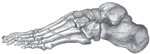
 Bones of the right foot. Dorsal surface.
 -
 Bones of the right foot. Plantar surface.
 -
 CT 3Dhuman Foot Skin and Bone
 -
 Skeleton of foot. Medial aspect.
 -
 Skeleton of foot. Lateral aspect.
 -
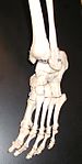 Bones of the feet from an actual skeleton.
 -
 Skeleton of Manus and Pes of a TailedBatrachian(from Professor Gegenbaur's "Tarsus and Carpus").
 -
 Bones of foot


See also
Notes
References
- Nordin, Margareta; Frankel, Victor Hirsch (2001). Basic biomechanics of the musculoskeletal system. Lippincott Williams & Wilkins. ISBN 0-683-30247-7.
- "Anatomy of the foot and ankle". Podiatry Channel. Archived from the original on 31 August 2009. Retrieved 30 August 2009.
- Romer, Alfred Sherwood; Parsons, Thomas S. (1977). The Vertebrate Body. Philadelphia, PA: Holt-Saunders International. pp. 205–208. ISBN 0-03-910284-X.
External links
- Diagram, identifying bones Archived 2016-03-04 at the Wayback Machine
- xrayslowerlimb at The Anatomy Lesson by Wesley Norman (Georgetown University) (xrayfootdorsal)