
Cerebrum
| Cerebrum | |
|---|---|
The lobes of the cerebral cortex include the frontal (blue), temporal (green), occipital (red), and parietal (yellow) lobes. The cerebellum (unlabeled) is not part of the telencephalon. | |
 Diagram depicting the main subdivisions of the embryonic vertebrate brain. | |
| Details | |
| Pronunciation | /ˈsɛrɪbrəm/, /sɪˈriːbrəm/ |
| Artery | anterior cerebral, middle cerebral, posterior cerebral |
| Vein | cerebral veins |
| Identifiers | |
| Latin | cerebrum |
| MeSH | D054022 |
| NeuroLex ID | birnlex_1042 |
| TA98 | A14.1.03.008 A14.1.09.001 |
| TA2 | 5416 |
| TH | H3.11.03.6.00001 |
| TE | E5.14.1.0.2.0.12 |
| FMA | 62000 |
| Anatomical terms of neuroanatomy | |

The cerebrum (pl.: cerebra), telencephalon or endbrain
With the assistance of the cerebellum, the cerebrum controls all voluntary actions in the human body.
Structure

The cerebrum is the largest part of the brain. Depending upon the position of the animal it lies either in front or on top of the brainstem. In humans, the cerebrum is the largest and best-developed of the five major divisions of the brain.
The cerebrum is made up of the two cerebral hemispheres and their
Cerebral cortex

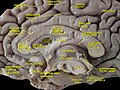
The cerebral cortex, the outer layer of grey matter of the cerebrum, is found only in mammals. In larger mammals, including humans, the surface of the cerebral cortex folds to create gyri (ridges) and sulci (furrows) which increase the surface area.[3]
The cerebral cortex is generally classified into four
Cerebral hemispheres
The cerebrum is divided by the
Development
In the developing vertebrate
The dorsal telencephalon then forms two lateral telencephalic vesicles, separated by the midline, which develop into the left and right cerebral hemispheres. Birds and fish have a dorsal telencephalon, like all vertebrates, but it is generally unlayered and therefore not considered a cerebral cortex. Only a layered cytoarchitecture can be considered a cortex.Functions
Note: As cerebrum is a gross division with many subdivisions and sub-regions, it is important to state that this section lists functions that cerebrum as a whole serves. See main articles on cerebral cortex and basal ganglia for more information. The cerebrum is a major part of the brain, controlling emotions, hearing, vision, personality and much more. It controls all precision of voluntary actions.
It functions as the center of sensory perception, memory, thoughts and judgement; the cerebrum also functions as the center of voluntary motor activities.
Sensory processing
The primary sensory areas of the cerebral cortex receive and process
Olfaction
The olfactory bulb, responsible for the sense of smell, takes up a large area of the cerebrum in most vertebrates. However, in humans, this part of the brain is much smaller and lies underneath the frontal lobe. The olfactory sensory system is unique since the neurons in the olfactory bulb send their axons directly to the olfactory cortex, rather than to the thalamus first. The olfaction is also the only sense that is represented by the ipsilateral side of the brain. Damage to the olfactory bulb results in a loss of olfaction (the sense of smell).
Language and communication
Learning and memory
Explicit or declarative (factual) memory formation is attributed to the
Implicit or procedural memory, such as complex motor behaviors, involves the basal ganglia.
Short-term or working memory involves association areas of the cortex, especially the dorsolateral prefrontal cortex, as well as the hippocampus.
Other animals
In the most primitive vertebrates, the
In
In the amniotes, the cerebrum becomes increasingly large and complex. In reptiles, the paleopallium is much larger than in amphibians and its growth has pushed the basal nuclei into the central regions of the cerebrum. As in the lower vertebrates, the grey matter is generally located beneath the white matter, but in some reptiles, it spreads out to the surface to form a primitive cortex, especially in the anterior part of the brain.[9]
In
The cerebra of
Additional images
-
 Cerebrum. Lateral face. Deep dissection.
 -
 Cerebrum. Medial face. Deep dissection.


See also
References
- ^ "BrainInfo". braininfo.rprc.washington.edu.
- ISBN 9780748736348. Retrieved 27 January 2015.
- ISBN 9780195028850. Retrieved 25 January 2015.
- ^ ISBN 9780781765213. Retrieved 28 January 2015.
- S2CID 7399128.
- S2CID 2406618.
- ISBN 978-0-87893-978-7.
- ISBN 978-0-07-139011-8.
- ^ ISBN 0-03-910284-X.
- ^ T.L. Brink (2008). "Unit 4: The Nervous System.". Psychology: A Student Friendly Approach (PDF). p. 62.
- PMID 15685220.
- PMID 16553307.