
Thalamus
| Thalamus | |
|---|---|
MRI cross-section) | |
 The thalamus in a 360° rotation | |
| Details | |
| Part of | Diencephalon |
| Parts | See List of thalamic nuclei |
| Artery | Posterior cerebral artery and branches |
| Identifiers | |
| Latin | thalamus dorsalis |
| MeSH | D013788 |
| NeuroNames | 300 |
| NeuroLex ID | birnlex_954 |
| TA98 | A14.1.08.101 A14.1.08.601 |
| TA2 | 5678 |
| TE | E5.14.3.4.2.1.8 |
| FMA | 62007 |
| Anatomical terms of neuroanatomy] | |
The thalamus (pl.: thalami; from
Anatomically, it is a paramedian symmetrical structure of two halves (left and right), within the
Anatomy
The thalamus is a paired structure of gray matter about four centimetres long, located in the forebrain which is superior to the midbrain, near the center of the brain with nerve fibers projecting out to the cerebral cortex in all directions. The medial surface of the thalamus constitutes the upper part of the lateral wall of the third ventricle, and is connected to the corresponding surface of the opposite thalamus by a flattened gray band, the interthalamic adhesion. The lateral part of the thalamus is the phylogenetically newest part of the thalamus (neothalamus), and includes the lateral nuclei, the pulvinar and the medial and lateral geniculate nuclei.[6][7] There are areas of white matter in the thalamus including the stratum zonale that covers the dorsal surface and the external and internal medullary laminae. The external lamina covers the lateral surface and the internal lamina divides the nuclei into anterior, medial, and lateral groups.[8]
Blood supply
The thalamus derives its blood supply from a number of arteries: the polar artery (posterior communicating artery), paramedian thalamic-subthalamic arteries, inferolateral (thalamogeniculate) arteries, and posterior (medial and lateral) choroidal arteries.[9] These are all branches of the posterior cerebral artery.[10]
Some people have the artery of Percheron, which is a rare anatomic variation in which a single arterial trunk arises from the posterior cerebral artery to supply both parts of the thalamus.
Thalamic nuclei


Derivatives of the
The thalamus comprises a system of
Connections

The thalamus has many connections to the hippocampus via the
The thalamus is connected to the cerebral cortex via the thalamocortical radiations.[14]
The
Function
The thalamus has multiple functions, and is generally believed to act as a relay station, or
The thalamus also plays an important role in regulating states of sleep and wakefulness.[18] Thalamic nuclei have strong reciprocal connections with the cerebral cortex, forming thalamo-cortico-thalamic circuits that are believed to be involved with consciousness.[19] The thalamus plays a major role in regulating arousal, the level of awareness, and activity. Damage to the thalamus can lead to permanent coma.[20]
The role of the thalamus in the more anterior
The neuronal information processes necessary for motor control were proposed as a network involving the thalamus as a subcortical motor center.[26] Through investigations of the anatomy of the brains of primates[27] the nature of the interconnected tissues of the cerebellum to the multiple motor cortices suggested that the thalamus fulfills a key function in providing the specific channels from the basal ganglia and cerebellum to the cortical motor areas.[28][29] In an investigation of the saccade and antisaccade[30] motor response in three monkeys, the thalamic regions were found to be involved in the generation of antisaccade eye-movement (that is, the ability to inhibit the reflexive jerking movement of the eyes in the direction of a presented stimulus).[31]
Recent research suggests that the mediodorsal thalamus (MD) may play a broader role in cognition. Specifically, the mediodorsal thalamus may "amplify the connectivity (signaling strength) of just the circuits in the cortex appropriate for the current context and thereby contribute to the flexibility (of the mammalian brain) to make complex decisions by wiring the many associations on which decisions depend into weakly connected cortical circuits."[32] Researchers found that "enhancing MD activity magnified the ability of mice to "think,"[32] driving down by more than 25 percent their error rate in deciding which conflicting sensory stimuli to follow to find the reward."[33]
Development
The thalamic complex is composed of the perithalamus (or prethalamus, previously also known as ventral thalamus), the mid-diencephalic organiser (which forms later the zona limitans intrathalamica (ZLI) ) and the thalamus (dorsal thalamus).[34][35] The development of the thalamus can be subdivided into three steps.[36] The thalamus is the largest structure deriving from the embryonic diencephalon, the posterior part of the forebrain situated between the midbrain and the cerebrum.
Early brain development
After
Formation of progenitor domains
Early in thalamic development two progenitor domains form, a caudal domain, and a rostral domain. The caudal domain gives rise to all of the glutamatergic neurons in the adult thalamus while the rostral domain gives rise to all of the GABAergic neurons in the adult thalamus.[41]
The formation of the mid-diencephalic organiser (MDO)
At the interface between the expression domains of Fez and Otx, the mid-diencephalic organizer (MDO, also called the ZLI organiser) is induced within the thalamic
Besides its importance as signalling center, the organizer matures into the morphological structure of the zona limitans intrathalamica (ZLI).
Maturation and parcellation of the thalamus
After its induction, the MDO starts to orchestrate the development of the thalamic anlage by release of signalling molecules such as SHH.[42] In mice, the function of signaling at the MDO has not been addressed directly due to a complete absence of the diencephalon in SHH mutants.[43]
Studies in chicks have shown that SHH is both necessary and sufficient for thalamic gene induction.[44] In zebrafish, it was shown that the expression of two SHH genes, SHH-a and SHH-b (formerly described as twhh) mark the MDO territory, and that SHH signaling is sufficient for the molecular differentiation of both the prethalamus and the thalamus but is not required for their maintenance and SHH signaling from the MDO/alar plate is sufficient for the maturation of prethalamic and thalamic territory while ventral Shh signals are dispensable.[45]
The exposure to SHH leads to differentiation of thalamic neurons. SHH signaling from the MDO induces a posterior-to-anterior wave of expression the proneural gene Neurogenin1 in the major (caudal) part of the thalamus, and Ascl1 (formerly Mash1) in the remaining narrow stripe of rostral thalamic cells immediately adjacent to the MDO, and in the prethalamus.[46][47]
This zonation of proneural gene expression leads to the differentiation of glutamatergic relay neurons from the Neurogenin1+ precursors and of GABAergic inhibitory neurons from the Ascl1+ precursors. In fish, selection of these alternative neurotransmitter fates is controlled by the dynamic expression of Her6 the homolog of
In humans, a common genetic variation in the promoter region of the
Clinical significance
A thalamus damaged by a
Atrophy of the thalamus is an indicator of MS.[52][53]
Additional images
-
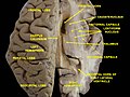 Human brain dissection, showing the thalamus.
 -
 Human thalamus along with other subcortical structures, in glass brain.
 -
 Lateral group of the thalamic nuclei.
 -
 Medial group of the thalamic nuclei.


See also
- 5-HT7 receptor
- Krista and Tatiana Hogan - conjoined twins with joined thalami
- List of regions in the human brain
- Nonmotor region of the ventral nuclear group of the thalamus
- Nucleus ventralis posterior lateralis pars oralis(VPLo), a region of the thalamus
- Primate basal ganglia system
- Thalamic stimulator
- Thalamotomy
References
- Retrieved 2012-02-09
- .
- ISBN 978-0-12-305460-9.
- ^ Gorvett, Zaria. "What you can learn from Einstein's quirky habits". bbc.com.
- S2CID 41337319.
- ^ "Medical Definition of NEOTHALAMUS". www.merriam-webster.com.
- ^ "neothalamus | Definition of neothalamus in English by Oxford Dictionaries". Oxford Dictionaries | English. Archived from the original on May 27, 2018.
- ISBN 978-0060466695.
- ^ Percheron, G. (1982). "The arterial supply of the thalamus". In Schaltenbrand; Walker, A. E. (eds.). Stereotaxy of the human brain. Stuttgart: Thieme. pp. 218–32.
- ^ Knipe, H Jones, J et al. Thalamus http://radiopaedia.org/articles/thalamus Archived 2017-09-17 at the Wayback Machine
- ^ Jones Edward G. (2007) "The Thalamus" Cambridge Uni. Press[page needed]
- ^ Percheron, G. (2003). "Thalamus". In Paxinos, G.; May, J. (eds.). The human nervous system (2nd ed.). Amsterdam: Elsevier. pp. 592–675.
- ^ S2CID 22002872.
- ^ University of Washington (1991). "Thalamocortical radiations". washington.edu.
- ISBN 978-0-393-91348-4.)
{{cite book}}: CS1 maint: multiple names: authors list (link - PMC 3055419.
- ^ "The thalamus, middleman of the brain, becomes a sensory conductor". The University of Chicago Medicine. Retrieved 10 September 2020.
- PMID 2839857.
- ISBN 978-1-447-12439-9p. 143
- ISBN 978-0-123-74168-4p. 10
- ^ Leonard, Abigail W. (August 17, 2006). "Your Brain Boots Up Like a Computer". LiveScience.
- PMID 11003270.
- S2CID 11258997.
- PMID 20550571.
- S2CID 11989085.
- PMID 4885774.
- S2CID 27155579.
- S2CID 25013002.
- PMID 15703228.
- ^ "The Antisaccade - A Review of Basic Research and Clinical Studies". Archived from the original on 2017-09-16. Retrieved 2012-02-10.[full citation needed]
- PMID 20371831.
- ^ a b "New Role Discovered For Brain Region". Neuroscience News. 2017-05-03. Retrieved 2017-12-03.
- PMID 28467827.
- S2CID 86730019.
- PMID 8575293.
- ^ PMID 20541814.
- PMID 16971467.
- PMID 17164418.
- PMID 9342056.
- PMID 17670791.
- PMID 25512300.
- S2CID 14658562.
- PMID 12361972.
- S2CID 29863625.
- PMID 16452095.
- PMID 19903880.
- PMID 19357274.
- S2CID 2214561.
- ^ Dejerine, J.; Roussy, G. (1906). "Le syndrome thalamique". Revue Neurologique. 14: 521–32.
- PMID 19151162.
- PMID 17416834.
- ^ Wexler, Marisa (20 October 2023). "Machine learning models estimate when brain atrophy starts in MS". Multiple Sclerosis News Today.
- PMC 10187410.