Thin section

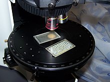
In
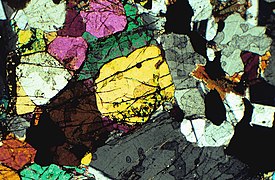
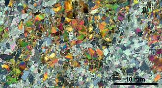
When placed between two polarizing filters set at right angles to each other, the optical properties of the minerals in the thin section alter the colour and intensity of the light as seen by the viewer. As different minerals have different optical properties, most rock forming minerals can be easily identified.
Thin sections are prepared in order to investigate the optical properties of the minerals in the rock. This work is a part of petrology and helps to reveal the origin and evolution of the parent rock.
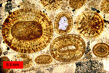
A photograph of a rock in thin section is often referred to as a
Thin sections are also used in the microscopic study of bones, metals and ceramics.
Quartz in thin section
Description
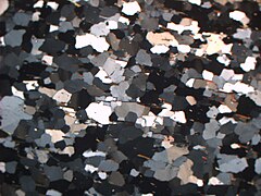
In thin section, when viewed in plane polarized light (PPL), quartz is colorless with low relief and no cleavage. Its habit is either fairly equant or anhedral if it infills around other minerals as a cement. Under cross polarized light (XPL) quartz displays low interference colors and is usually the defining mineral used to determine if the thin section is at standardized thickness of 30 microns as quartz will only display up to a very pale yellow interference color and no further at that thickness, and it is very common in most rocks so it will likely be available to judge the thickness.[1]
Determining provenance
In thin section, quartz grain
Other distinguishing features
The above descriptions of quartz in thin section are usually enough to identify it. Minerals with similar appearance may include

Ultra-thin sections
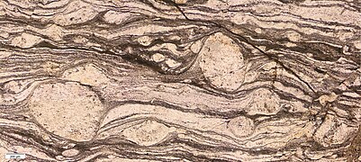
Fine-grained rocks, particularly those containing minerals of high
Gallery
See also
- Ceramography: thin sections of ceramics
References
- ^ "Rock Thin Sections (Petrographic Thin Section Preparation)". Kemet. Retrieved 2018-05-15.
- OCLC 946008550.
- ^ Blatt, H.; Christie, J.M. (1963). "Undulatory Extinction in Quartz of Igneous and Metamorphic Rocks and Its Significance in Provenance Studies of Sedimentary Rocks". AAPG Datapages.
- )
- ^ "quartz". www.mtholyoke.edu. Retrieved 2018-05-15.
- ^ Badertscher, N.P. & Burkhard, M. 2000. Brittle±ductile deformation in the Glarus thrust Lochseiten (LK) calc-mylonite, Terra Nova, 12, 281-288
- ^ Barber, D.J. 1981. Demountable polished extra-thin sections and their use in transmission electron microscopy. Mineralogical magazine,44, 357-359
- Shelley, D. Optical Mineralogy, Second Edition. University of Canterbury, New Zealand.
External links
- Thin sections of soils. Collection of Prof. Kubiëna Archived 22 September 2020 at the Wayback Machine
- Uncommon igneous, metamorphic and metasomatic rocks in thin section, in unpolarized light and under crossed polarizers
- Namethatmineral.com: dynamic data-tables for identification of thin sections under the microscope

