
Tibia
| tibiae | |
|---|---|
superior and inferior tibiofibular joint | |
| Identifiers | |
| Latin | (os) tibia |
| MeSH | D013977 |
| TA98 | A02.5.06.001 |
| TA2 | 1397 |
| FMA | 24476 |
| Anatomical terms of bone] | |
The tibia (
Structure
In
The
The tibia is categorized as a
Upper extremity
Condyles of tibia


The proximal or upper extremity of the tibia is expanded in the transverse plane with a
The medial and lateral condyle are separated by the
Facets
The superior articular surface presents two smooth articular facets.
- The medial facet, oval in shape, is slightly concave from side to side, and from before backward.
- The lateral, nearly circular, is concave from side to side, but slightly convex from before backward, especially at its posterior part, where it is prolonged on to the posterior surface for a short distance.
The central portions of these facets articulate with the condyles of the femur, while their peripheral portions support the menisci of the knee joint, which here intervene between the two bones.
Intercondyloid eminence
Between the articular facets in the intercondylar area, but nearer the posterior than the anterior aspect of the bone, is the intercondyloid eminence (spine of tibia), surmounted on either side by a prominent tubercle, on to the sides of which the articular facets are prolonged; in front of and behind the intercondyloid eminence are rough depressions for the attachment of the anterior and posterior cruciate ligaments and the menisci.
Surfaces
The anterior surfaces of the condyles are continuous with one another, forming a large somewhat flattened area; this area is triangular, broad above, and perforated by large vascular foramina; narrow below where it ends in a large oblong elevation, the tuberosity of the tibia, which gives attachment to the
Posteriorly, the condyles are separated from each other by a shallow depression, the posterior intercondyloid fossa, which gives attachment to part of the
Its medial surface is convex, rough, and prominent; it gives attachment to the medial collateral ligament.
The lateral condyle presents posteriorly a flat articular facet, nearly circular in form, directed downward, backward, and lateralward, for articulation with the head of the fibula. Its lateral surface is convex, rough, and prominent in front: on it is an eminence, situated on a level with the upper border of the tuberosity and at the junction of its anterior and lateral surfaces, for the attachment of the
Shaft

The shaft or body of the tibia is triangular in cross-section and forms three borders: an anterior, medial, and lateral or interosseous border. These three borders form three surfaces: the medial, lateral, and posterior.[2] The forward flat part of the tibia is called the fibia, often confused with the fibula.[3][failed verification]
Borders
The anterior crest or border, the most prominent of the three, commences above at the tuberosity, and ends below at the anterior margin of the medial malleolus. It is sinuous and prominent in the upper two-thirds of its extent, but smooth and rounded below; it gives attachment to the deep fascia of the leg.
The medial border is smooth and rounded above and below, but more prominent in the center. It begins at the back part of the medial condyle, and ends at the posterior border of the medial malleolus; its upper part gives attachment to the tibial collateral ligament of the knee-joint to the extent of about 5 cm., and insertion to some fibers of the
The interosseous crest or lateral border is thin and prominent, especially its central part, and gives attachment to the interosseous membrane; it commences above in front of the fibular articular facet, and bifurcates below, to form the boundaries of a triangular rough surface, for the attachment of the interosseous ligament connecting the tibia and fibula.
Surfaces
The medial surface is smooth, convex, and broader above than below; its upper third, directed forward and medialward, is covered by the
The lateral surface is narrower than the medial; its upper two-thirds present a shallow groove for the origin of the Tibialis anterior; its lower third is smooth, convex, curves gradually forward to the anterior aspect of the bone, and is covered by the tendons of the
The posterior surface presents, at its upper part, a prominent ridge, the popliteal line, which extends obliquely downward from the back part of the articular facet for the fibula to the medial border, at the junction of its upper and middle thirds; it marks the lower limit of the insertion of the
Lower extremity
.jpg)
.jpg)
The distal end of the tibia is much smaller than the proximal end and presents five surfaces; it is prolonged downward on its medial side as a strong pyramidal process, the medial malleolus. The lower extremity of the tibia together with the fibula and talus forms the ankle joint.
Surfaces
The inferior articular surface is quadrilateral, and smooth for articulation with the talus. It is concave from before backward, broader in front than behind, and traversed from before backward by a slight elevation, separating two depressions. It is continuous with that on the medial malleolus.
The anterior surface of the lower extremity is smooth and rounded above, and covered by the tendons of the Extensor muscles; its lower margin presents a rough transverse depression for the attachment of the articular capsule of the ankle-joint.
The posterior surface is traversed by a shallow groove directed obliquely downward and medialward, continuous with a similar groove on the posterior surface of the talus and serving for the passage of the tendon of the
The lateral surface presents a triangular rough depression for the attachment of the inferior interosseous ligament connecting it with the fibula; the lower part of this depression is smooth, covered with cartilage in the fresh state, and articulates with the fibula. The surface is bounded by two prominent borders (the anterior and posterior colliculi), continuous above with the interosseous crest; they afford attachment to the anterior and posterior ligaments of the lateral malleolus.
The medial surface -- see
Fractures
Ankle fractures of the tibia have several classification systems based on location or mechanism:
- Medial malleolus - Herscovici classification
- Haruguchi classification
- Mechanism - Lauge-Hansen classification
Blood supply
The tibia is supplied with blood from two sources: A nutrient artery, as the main source, and periosteal vessels derived from the anterior tibial artery.[4]
Joints
The tibia is a part of four joints; the knee, ankle,
In the knee the tibia forms one of the two
The part of the ankle joint known as the talocrural joint, is a synovial hinge joint that connects the distal ends of the tibia and fibula in the lower limb with the proximal end of the talus. The articulation between the tibia and the talus bears more weight than between the smaller fibula and the talus.[citation needed]


Development
The tibia is
The center for the upper epiphysis appears before or shortly after birth at close to 34 weeks gestation; it is flattened in form, and has a thin tongue-shaped process in front, which forms the
The lower epiphysis fuses with the tibial shaft at about the eighteenth, and the upper one fuses about the twentieth year.
Two additional centers occasionally exist, one for the tongue-shaped process of the upper epiphysis, which forms the tuberosity, and one for the
Function
Muscle attachments
| Muscle | Direction | Attachment[7] |
| Tensor fasciae latae muscle | Insertion | Gerdy's tubercle |
Quadriceps femoris muscle |
Insertion | Tuberosity of the tibia |
| Sartorius muscle | Insertion | Pes anserinus |
| Gracilis muscle | Insertion | Pes anserinus |
| Semitendinosus muscle | Insertion | Pes anserinus |
| Horizontal head of the semimembranosus muscle | Insertion | Medial condyle |
| Popliteus muscle | Insertion | Posterior side of the tibia over the soleal line |
| Tibialis anterior muscle | Origin | Lateral side of the tibia |
| Extensor digitorum longus muscle | Origin | Lateral condyle |
| Soleus muscle | Origin | Posterior side of the tibia under the soleal line |
| Flexor digitorum longus muscle | Origin | Posterior side of the tibia under the soleal line |
Strength
The tibia has been modeled as taking an axial force during walking that is up to 4.7 bodyweight. Its bending moment in the sagittal plane in the late stance phase is up to 71.6 bodyweight times millimetre.[8]
Clinical significance
Fracture
Society and culture
In Judaism, the tibia, or shankbone, of a goat or sheep is used in the Passover Seder plate.
Other animals
The structure of the tibia in most other
Additional images
-
 Shape of right tibia
 -
 3D image
 -
 Longitudinal section of tibia showing interior
 -
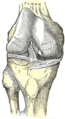
 Right knee-joint. Anterior view.
 -
 Right knee joint from the front, showing interior ligaments
 -
 Left knee joint from behind, showing interior ligaments
 -
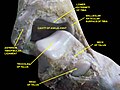
 Left talocrural joint
 -
 Coronal section through right talocrural and talocalcaneal joints
 -
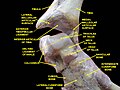
 Dorsum of Foot. Ankle joint. Deep dissection
 -
 Dorsum of Foot. Ankle joint. Deep dissection
 -
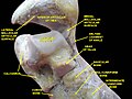
 Ankle joint. Deep dissection. Anterior view
 -
 Bones of the right leg. Anterior surface
 -
 Bones of the right leg. Posterior surface
 -
 Dorsum of Foot. Ankle joint. Deep dissection.
 -
 Ankle joint. Deep dissection.
 -
 Ankle joint. Deep dissection.
 -
 Ankle joint. Deep dissection.
 -
 Ankle joint. Deep dissection.
 -
Tibia Anatomy


See also
- Shin splints
- Squatting facets
References
![]() This article incorporates text in the public domain from page 256 of the 20th edition of Gray's Anatomy (1918)
This article incorporates text in the public domain from page 256 of the 20th edition of Gray's Anatomy (1918)
- ^ ISBN 978-0-443-06952-9.[page needed]
- ^ ISBN 978-0-443-06952-9.
- ^ "Chapter 12: THE BONES OF THE LOWER LIMB". www.dartmouth.edu. Retrieved 2018-06-17.
- PMID 13854090.
- PMID 19594940.
- PMID 19726621.
- ISBN 978-87-628-0307-7.
- PMID 19185959.
- ISBN 0-03-910284-X.