
Tibial nerve
| Tibial nerve | |
|---|---|
abductor digiti minimi | |
| Identifiers | |
| Latin | nervus tibialis |
| MeSH | D013979 |
| TA98 | A14.2.07.058 |
| TA2 | 6582 |
| FMA | 19035 |
| Anatomical terms of neuroanatomy] | |
The tibial nerve is a branch of the sciatic nerve. The tibial nerve passes through the popliteal fossa to pass below the arch of soleus.
Structure
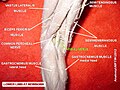
Popliteal fossa
The tibial nerve is the larger terminal branch of the sciatic nerve with root values of L4, L5, S1, S2, and S3. It lies superficial (or posterior) to the popliteal vessels, extending from the superior angle to the inferior angle of the popliteal fossa, crossing the popliteal vessels from lateral to medial side. It gives off branches as shown below:[1]
- Muscular branches - Muscular branches arise from the distal part of the popliteal fossa. It supplies the medial and lateral heads of tibialis posterior muscle, superior tibiofibular joint, tibia bone, interosseous membrane of leg, and the inferior tibiofibular joint.[1]
- Cutaneous branches - Tibial nerve also gives off a cutaneous nerve called the medial sural cutaneous nerve from the middle of the popliteal fossa and exits at the inferior angle. It supplies the skin of the lower half of the back of the leg and lateral border of the foot until the tip of the little toe.[1]
- Articular branches - There are three articular branches arises from the upper part of the fossa: superior medial genicular nerve (located on the surface of medial condyle of femur, middle genicular nerve (pierces the posterior capsule of the knee joint to supply the structures located in the intercondylar notch of the femur, and inferior genicular nerve (runs along the upper border of the popliteus to reach the medial condyle of tibia).[1]
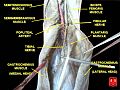
Back of the leg
At the inferior angle of the popliteal fossa, tibial nerve passes deep to the tendinous arch of soleus to enter the back of the leg. In the leg, it runs downwards and medially to reach the posteromedial side of the ankle, midway between the
- Muscular branches - Supplies tibialis posterior, flexor hallucis longus, and deep part of soleus.[1]
- Cutaneus branches - The medial calcaneal nerve pierces the flexor retinaculum to supply the skin of the back and lower surface of the heel.[1]
- Articular branches - Supplies the ankle joint[1]
Foot
In the foot, the nerve terminates by dividing into medial and lateral plantar branches.
- flexor hallucis brevis and the first lumbrical. Cutaneous distribution of the medial plantar nerve supplies the medial sole and medial three and one half toes through four digital branches. Each digital branch give off a dorsal branch to supply the nail beds on the dorsum. This nerve also gives off articular branches to supply the bones of the tarsus and metatarsus.[1]
- adductor hallucis.[1]
Clinical significance
This section includes a improve this section by introducing more precise citations. (October 2023) ) |
Damage to the tibial nerve is rare, and is often a result of direct trauma, entrapment through narrow space or compression for long period of time. Damage results in loss of plantar flexion, loss of flexion of toes and weakened inversion (The tibialis anterior can still invert the foot).
Additional images
-
 Tibial nerve
 -
 Cross-section through middle of left calf
 -
 Cutaneous nerves of the right lower extremity. Front and posterior views
 -
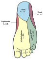 Diagram of the segmental distribution of the cutaneous nerves of the sole of the foot
 -
 A schematic of the sacral plexus with the origin of the tibial nerve shown (labeled at the bottom left)
 -
 Tibial nerve
 -
 Tibial nerve
 -
 Tibial nerve


References
- ^ ISBN 978-81-239-1864-8.
External links
- Tibial nerve at the Duke University Health System's Orthopedics program