
Dermatophytosis
| Dermatophytosis | |
|---|---|
| Other names | Ringworm, tinea |
| Frequency | 20% of the population[8] |
Dermatophytosis, also known as ringworm, is a
About 40 types of fungus can cause ringworm.
Prevention is by keeping the skin dry, not walking barefoot in public, and not sharing personal items.
Globally, up to 20% of the population may be infected by ringworm at any given time.[8] Infections of the groin are more common in males, while infections of the scalp and body occur equally in both sexes.[4] Infections of the scalp are most common in children while infections of the groin are most common in the elderly.[4] Descriptions of ringworm date back to ancient history.[9]
Signs and symptoms
Animals including dogs and cats can also be affected by ringworm, and the disease can be transmitted between animals and humans, making it a
Specific signs can be:
- red, scaly, itchy or raised patches
- patches may be redder on outside edges or resemble a ring
- patches that begin to ooze or develop a blister
- bald patches may develop when the scalp is affected
Causes
Diagnosis
Dermatophyte infections can be readily diagnosed based on the history, physical examination, and potassium hydroxide (KOH) microscopy.[10]
Classification

A number of different species of fungus are involved in dermatophytosis.
- Dermatophytosis
- Tinea pedis (athlete's foot): fungal infection of the feet
- toenails, and the nail bed
- Tinea corporis: fungal infection of the arms, legs, and trunk
- jock itch): fungal infection of the groin area
- handsand palm area
- Tinea capitis: fungal infection of the scalp and hair
- Tinea faciei (face fungus): fungal infection of the face
- Tinea barbae: fungal infestation of facial hair
- Other superficial mycoses (not classic ringworm, since not caused by dermatophytes)
- Tinea versicolor: caused by Malassezia furfur
- Tinea nigra: caused by Hortaea werneckii
Prevention
Advice often given includes:
- Avoid sharing clothing, sports equipment, towels, or sheets.
- Wash clothes in hot water with fungicidalsoap after suspected exposure to ringworm.
- Avoid walking barefoot; instead wear appropriate protective shoes in locker rooms and sandals at the beach.[11][12][13]
- Avoid touching pets with bald spots, as they are often carriers of the fungus.
Vaccination
As of 2016,[update] no approved human vaccine exist against dermatophytosis. For
Treatment
In more severe cases or scalp ringworm,
To prevent spreading the infection, lesions should not be touched, and good hygiene maintained with washing of hands and the body.[22]
Misdiagnosis and treatment of ringworm with a
History
Dermatophytosis has been prevalent since before 1906, at which time ringworm was treated with compounds of
Terminology
The most common term for the infection, "ringworm", is a
Other animals
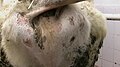
Ringworm caused by Trichophyton verrucosum is a frequent clinical condition in cattle. Young animals are more frequently affected. The lesions are located on the head, neck, tail, and perineum.[25] The typical lesion is a round, whitish crust. Multiple lesions may coalesce in "map-like" appearance.
-
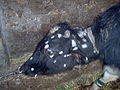 Multiple lesions, head
 -
 Around the eyes and on ears
 -
 On cheeks: crusted lesion (right)
 -
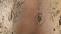 Old lesions, with regrowing hair
 -
 On neck and withers
 -
 On perineum


Clinical dermatophytosis is also diagnosed in sheep, dogs, cats, and horses. Causative agents, besides Trichophyton verrucosum, are T. mentagrophytes, T. equinum, Microsporum gypseum, M. canis, and M. nanum.[26]
Dermatophytosis may also be present in the
Diagnosis
Ringworm in pets may often be asymptomatic, resulting in a
Veterinarians have several tests to identify ringworm infection and identify the fungal species that cause it:
Woods test: This is an
Microscopic test: The veterinarian takes hairs from around the infected area and places them in a staining solution to view under the microscope. Fungal spores may be viewed directly on hair shafts. This technique identifies a fungal infection in about 40%–70% of the infections, but cannot identify the species of dermatophyte.
Culture test: This is the most effective, but also the most time-consuming, way to determine if ringworm is on a pet. In this test, the veterinarian collects hairs from the pet, or else collects fungal spores from the pet's hair with a toothbrush, or other instrument, and inoculates fungal media for culture. These cultures can be brushed with transparent tape and then read by the veterinarian using a microscope, or can be sent to a pathological lab. The three common types of fungi which commonly cause pet ringworm can be identified by their characteristic spores. These are different-appearing
Identifying the species of fungi involved in pet infections can be helpful in controlling the source of infection. M. canis, despite its name, occurs more commonly in domestic cats, and 98% of cat infections are with this organism.[
Treatment
Pet animals
Treatment requires both systemic oral treatment with most of the same drugs used in humans—terbinafine, fluconazole, or itraconazole—as well as a topical "dip" therapy.[28]
Because of the usually longer hair shafts in pets compared to those of humans, the area of infection and possibly all of the longer hair of the pet must be clipped to decrease the load of fungal spores clinging to the pet's hair shafts. However, close shaving is usually not done because nicking the skin facilitates further skin infection.
Twice-weekly bathing of the pet with diluted lime sulfur dip solution is effective in eradicating fungal spores. This must continue for 3 to 8 weeks.[29]
Washing of household hard surfaces with 1:10 household sodium hypochlorite bleach solution is effective in killing spores, but it is too irritating to be used directly on hair and skin.
Pet hair must be rigorously removed from all household surfaces, and then the vacuum cleaner bag, and perhaps even the vacuum cleaner itself, discarded when this has been done repeatedly. Removal of all hair is important, since spores may survive 12 months or even as long as two years on hair clinging to surfaces.[30]
Cattle
In
Epidemiology
Worldwide, superficial fungal infections caused by dermatophytes are estimated to infect around 20-25% of the population and it is thought that dermatophytes infect 10-15% of the population during their lifetime.[31][32] The highest incidence of superficial mycoses result from dermatophytoses which are most prevalent in tropical regions.[31][33] Onychomycosis, a common infection caused by dermatophytes, is found with varying prevalence rates in many countries.[34] Tinea pedis + onychomycosis, Tinea corporis, Tinea capitis are the most common dermatophytosis found in humans across the world.[34] Tinea capitis has a greater prevalence in children.[31] The increasing prevalence of dermatophytes resulting in Tinea capitis has been causing epidemics throughout Europe and America.[34] In pets, cats are the most affected by dermatophytosis.[35] Pets are susceptible to dermatophytoses caused by Microsporum canis, Microsporum gypseum, and Trichophyton.[35] For dermatophytosis in animals, risk factors depend on age, species, breed, underlying conditions, stress, grooming, and injuries.[35]
Numerous studies have found Tinea capitis to be the most prevalent dermatophyte to infect children across the continent of Africa.[32] Dermatophytosis has been found to be most prevalent in children ages 4 to 11, infecting more males than females.[32] Low socioeconomic status was found to be a risk factor for Tinea capitis.[32] Throughout Africa, dermatophytoses are common in hot- humid climates and with areas of overpopulation.[32]
Chronicity is a common outcome for dermatophytosis in India.[33] The prevalence of dermatophytosis in India is between 36.6 and 78.4% depending on the area, clinical subtype, and dermatophyte isolate.[33] Individuals ages 21–40 years are most commonly affected.[33]
A 2002 study looking at 445 samples of dermatophytes in patients in Goiânia, Brazil found the most prevalent type to be Trichophyton rubrum (49.4%), followed by Trichophyton mentagrophytes (30.8%), and Microsporum canis (12.6%).[36]
A 2013 study looking at 5,175 samples of Tinea in patients in Tehran, Iran found the most prevalent type to be Tinea pedis (43.4%), followed by Tinea unguium. (21.3%), and Tinea cruris (20.7%).[37]
See also
- fungipresent in a particular niche like the human body.
- Lichen planus - an autoimmune disease that produces similar skin blotching to ringworm.
References
- ^ a b c d "Symptoms of Ringworm Infections". CDC. December 6, 2015. Archived from the original on 20 January 2016. Retrieved 5 September 2016.
- ^ a b c d "Definition of Ringworm". CDC. December 6, 2015. Archived from the original on 5 September 2016. Retrieved 5 September 2016.
- ^ a b c d e "Ringworm Risk & Prevention". CDC. December 6, 2015. Archived from the original on 7 September 2016. Retrieved 5 September 2016.
- ^ ISBN 9781451188509. Archivedfrom the original on 2016-09-15.
- ^ a b c "Diagnosis of Ringworm". CDC. December 6, 2015. Archived from the original on 8 August 2016. Retrieved 5 September 2016.
- ISBN 9780781770453. Archivedfrom the original on 2017-04-26.
- ^ a b c "Treatment for Ringworm". CDC. December 6, 2015. Archived from the original on 3 September 2016. Retrieved 5 September 2016.
- ^ ISBN 978-0-8493-8786-9. Archivedfrom the original on 8 September 2017.
- ISBN 978-0702051821. Archivedfrom the original on 2016-09-15.
- PMID 12537173.)
{{cite journal}}: CS1 maint: multiple names: authors list (link - ^ Klemm L (2 April 2008). "Keeping footloose on trips". The Herald News. Archived from the original on 18 February 2009.
- ^ Fort Dodge Animal Health: Milestones from Wyeth.com. Retrieved April 28, 2008.
- ^ "Ringworm In Your Dog, Cat And Other Pets". Vetspace. Retrieved 14 November 2020.
- ^ "Insol Dermatophyton 5x2 ml". GROVET - The veterinary warehouse. Archived from the original on 2016-08-17. Retrieved 2016-02-01.
- PMID 12603775
- ^ S2CID 37500893.
- S2CID 195691703.
- ^ Tinea~treatment at eMedicine
- ^ Tinea Corporis~treatment at eMedicine
- PMID 19145261.
- S2CID 24116721.
- ^ "Ringworm on Body Treatment" at eMedicineHealth
- (PDF) from the original on 2009-11-22.
- ^ Mrs. M. Grieve. A Modern Herbal. Archived from the original on 2015-03-25.
- ISBN 978-0-8138-0516-0.
- ^ a b David W. Scott, Colour Atlas of Animal Dermatology, Blackwell Publishing Professional 2121 State Avenue, Ames, Iowa 50014, USA; ISBN 978-0-8138-0516-0/2007.
- ^ "General ringworm information". Ringworm.com.au. Archived from the original on 2010-12-21. Retrieved 2011-01-10.
- ^ "Facts About Ringworm". Archived from the original on 2011-10-06. Retrieved 2011-10-03. Detailed veterinary discussion of animal treatment
- ^ "Veterinary treatment site page". Marvistavet.com. Archived from the original on 2013-01-04. Retrieved 2011-01-10.
- ^ "Persistence of spores". Ringworm.com.au. Archived from the original on 2010-12-21. Retrieved 2011-01-10.
- ^ a b c Pires, C. A. A., Cruz, N. F. S. da, Lobato, A. M., Sousa, P. O. de, Carneiro, F. R. O., & Mendes, A. M. D. (2014). Clinical, epidemiological, and therapeutic profile of dermatophytosis. Anais Brasileiros de Dermatología, 89(2), 259–264. [1]
- ^ a b c d e Oumar Coulibaly, Coralie L'Ollivier, Renaud Piarroux, Stéphane Ranque, Epidemiology of human dermatophytoses in Africa, Medical Mycology, Volume 56, Issue 2, February 2018, Pages 145–161.
- ^ a b c d Rajagopalan, M., Inamadar, A., Mittal, A., Miskeen, A. K., Srinivas, C. R., Sardana, K., Godse, K., Patel, K., Rengasamy, M., Rudramurthy, S., & Dogra, S. (2018). Expert Consensus on The Management of Dermatophytosis in India (ECTODERM India). BMC dermatology, 18(1), 6. [2]
- ^ a b c Hayette, M.-P., & Sacheli, R. (2015). Dermatophytosis, Trends in Epidemiology and Diagnostic Approach. Current Fungal Infection Reports, 9(3), 164–179. [3]
- ^ a b c Gordon, E., Idle, A., & DeTar, L. (2020). Descriptive epidemiology of companion animal dermatophytosis in a Canadian Pacific Northwest animal shelter system. The Canadian veterinary journal = La revue veterinaire canadienne, 61(7), 763–770.
- ^ Costa, M., Passos, X. S., Hasimoto e Souza, L. K., Miranda, A. T. B., Lemos, J. de A., Oliveira, J., & Silva, M. do R. R. (2002). Epidemiology and etiology of dermatophytosis in Goiânia, GO, Brazil. Revista da Sociedade Brasileira de Medicina Tropical, 35(1), 19–.
- ^ Rezaei-Matehkolaei, A., Makimura, K., de Hoog, S., Shidfar, M. R., Zaini, F., Eshraghian, M., Naghan, P. A., & Mirhendi, H. (2013). Molecular epidemiology of dermatophytosis in Tehran, Iran, a clinical and microbial survey. Medical Mycology (Oxford), 51(2), 203–207. [4]
Further reading
- Weitzman I, Summerbell RC (1995). "The dermatophytes". Clinical Microbiology Reviews. 8 (2): 240–259. PMID 7621400.
- Pietro Nenoff, Constanze Krüger, Gabriele Ginter-Hanselmayer, Hans-Jürgen Tietz (2014) Mycology – an update. Part 1: Dermatomycoses: Causative agents, epidemiology and pathogenesis
External links
- Tinea photo library at Dermnet Archived 2008-10-15 at the Wayback Machine
| Authority control databases: National |
|---|