
Trachea
| Trachea | |
|---|---|
tracheal branches of inferior thyroid artery | |
| Vein | brachiocephalic vein, azygos vein accessory hemiazygos vein |
| Identifiers | |
| Latin | trachea |
| MeSH | D014132 |
| TA98 | A06.3.01.001 |
| TA2 | 3213 |
| FMA | 7394 |
| Anatomical terminology] | |
The trachea (pl.: tracheae or tracheas), also known as the windpipe, is a
The trachea begins to form in the second month of embryo development, becoming longer and more fixed in its position over time. It is
The word trachea is used to define a very different organ in
Structure

An adult's trachea has an inner diameter of about 1.5 to 2 centimetres (1⁄2 to 3⁄4 in) and a length of about 10 to 11 cm (4 to 4+1⁄4 in), wider in males than females.
Although trachea is a midline structure, it can be displaced normally to the right by the aortic arch.[5]
Nearby structures
The trachea passes by many structures of the neck and chest (thorax) along its course.
In front of the upper trachea lies connective tissue and skin.
Behind the trachea, along its length, sits the
The trachealis muscle contracts during coughing, reducing the size of the lumen of the trachea.[3]
-
CT scan of the thorax (axial lung window)
-
CT scan of the thorax (coronal lung window)
-
CT scan of the thorax (coronal mediastinal window)
-
 Cross section of a trachea and esophagus
 -
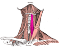 The sternohyoid and sternothyroid muscles lie on top of the upper part of the trachea
 -
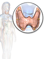 The thyroid gland also lies on top of the trachea, and lies below the cricoid cartilage.


Blood and lymphatic supply

The upper part of trachea receives and drains
Development
In the fourth week of
The trachea is no more than 4 mm in diameter during the first year of life, expanding to its adult diameter of approximately 2cm by late childhood.[2][3] The trachea is more circular and more vertical in children compared to adults,[3] varies more in size, and also varies more in its position in relation to its surrounding structures.[2]
Microanatomy
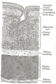
The trachea is lined with a layer of
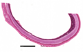
The trachea is surrounded by 16 to 20 rings of hyaline cartilage; these 'rings' are incomplete and C-shaped.[2] Two or more of the cartilages often unite, partially or completely, and they are sometimes bifurcated at their extremities. The rings are generally highly elastic but they may calcify with age.
-
 Cross-section
 -
 Cross-section of the trachea, with pseudostratified ciliated columnar epithelium and goblet cells labelled
 -
 Magnified cross-section of the cartilage of the trachea


Function
The trachea's main function is to transport air to and from the lungs. It also helps to warm, humidify, and filter the air before it reaches the lungs.
The trachea is made up of rings of cartilage, which help to keep it open and prevent it from collapsing. The inside of the trachea is lined with a mucous membrane, which produces mucus to help trap dirt and dust particles. The cilia, which are tiny hairs that line the mucous membrane, help to move the mucus and trapped particles up and out of the trachea.
Clinical significance
Inflammation and infection
A person affected with tracheitis may start with symptoms that suggest an
Narrowing
A trachea may be
One cause of narrowing is
Injury
The trachea may be injured by trauma such as in a vehicle accident, or intentionally by another wilfully inflicting damage for example as practiced in some martial arts.[16]
Intubation
Tracheal intubation refers to the insertion of a
In an emergency, or when tracheal intubation is deemed impossible, a
Congenital disorders

Tracheal agenesis[20] is a rare birth defect in which the trachea fails to develop. The defect is usually fatal though sometimes surgical intervention has been successful.
A
Sometimes as an anatomical variation one or more of the tracheal rings are formed as complete rings, rather than horseshoe shaped rings. These O rings are smaller than the normal C-shaped rings and can cause narrowing (stenosis) of the trachea, resulting in breathing difficulties. An operation called a slide tracheoplasty can open up the rings and rejoin them as wider rings, shortening the length of the trachea.[22] Slide tracheoplasty is said to be the best option in treating tracheal stenosis.[23]
Replacement
From 2008, operations have experimentally replaced tracheas, with those grown from stem cells, or with synthetic substitutes, however this is regarded as experimental and there is no standardised method.[25] Difficulties with ensuring adequate blood supply to the replaced trachea is considered a major challenge to any replacement. Additionally, no evidence has been found to support the placement of stem cells taken from bone marrow on the trachea as a way of stimulating tissue regeneration, and such a method remains hypothetical.[25]
In January 2021, surgeons at Mount Sinai Hospital in New York performed the first complete trachea transplantation. The 18-hour procedure included harvesting a trachea from a donor and implanting it in the patient, connecting numerous veins and arteries to provide sufficient blood flow to the organ.[26]
Other animals
Allowing for variations in the length of the neck, the trachea in other mammals is, in general, similar to that in humans. Generally, it is also similar to the reptilian trachea.[27]
Vertebrates
In
In amphibians, the trachea is normally extremely short, and leads directly into the lungs, without clear primary bronchi. A longer trachea is, however, found in some long-necked salamanders, and in caecilians. While there are irregular cartilagenous nodules on the amphibian trachea, these do not form the rings found in amniotes.[27]
The only vertebrates to have lungs, but no trachea, are the lungfish and the Polypterus, in which the lungs arise directly from the pharynx.[27]
Invertebrates

The word trachea is used to define a very different organ in
A tracheal tube may contain ridge-like circumferential rings of taenidia in various geometries such as loops or helices. Taenidia provide strength and flexibility to the trachea. In the head, thorax, or abdomen, tracheae may also be connected to air sacs. Many insects, such as grasshoppers and bees, which actively pump the air sacs in their abdomen, are able to control the flow of air through their body. In some aquatic insects, the tracheae exchange gas through the body wall directly, in the form of a gill, or function essentially as normal, via a plastron. Note that despite being internal, the tracheae of arthropods are lined with cuticular tissue and are shed during moulting (ecdysis).[29]
Additional images
-
 Trachea (mammal) cross-section high resolution
 -
 Trachea (mammal) cross-section low resolution
 -
 Trachea
 -
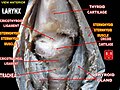
 Coronal section of larynx and upper part of trachea
_histology_cross-section.png)
_histology_cross-section_low_mag.png)


References
- ^ "Trachea | Definition of Trachea by Lexico". Lexico Dictionaries | English. Archived from the original on 7 July 2020. Retrieved 27 October 2019.
- ^ )
- ^ PMID 29707503.
- PMID 9423323.
- PMID 8462036.
- ^ ISBN 9781496383907.
- ISBN 978-0-07-163020-7. Archived from the originalon 3 June 2013. Retrieved 24 February 2015.
- S2CID 9551913.
- ^ S2CID 86826724.
- ^ S2CID 42828725.
- ^ Bhatia R. "Bacterial Tracheitis - Pediatrics". Merck Manuals Professional Edition. Retrieved 21 May 2020.
- ^ ISBN 978-0-7020-7028-0.
- ^ PMID 25962857.
- PMID 20051001.
- PMID 24268337.
- PMID 29763191. Retrieved 23 October 2022.
- ^ "Definition of INTUBATION". www.merriam-webster.com. Merriam Webster. Retrieved 25 May 2020.
- ^ Molnar H (11 April 2023). "Types of Tracheostomy Tubes".
- ^ "Medical Definition of CRICOTHYROTOMY". www.merriam-webster.com. Merriam Webster. Retrieved 25 May 2020.
- S2CID 21260092.
- ^ "Evaluation and Treatment for Tracheoesophageal Puncture and Prosthesis: Technical Report". American Speech-Language-Hearing Association. American Speech-Language-Hearing Association (ASHA). 2004. Retrieved 26 May 2020.
- ^ "Slide tracheoplasty". Retrieved 2 October 2015.
- S2CID 25335958.
- PMID 18176298.
- ^ PMID 26981270.
- ^ Harris R (6 April 2021). "Woman Gets New Windpipe In Groundbreaking Transplant Surgery". NPR. Retrieved 6 April 2021.
- ^ ISBN 978-0-03-910284-5.
- ISBN 0-471-15955-7.
- ^ OCLC 55793895.
- S2CID 43634044.
| National | |
|---|---|
| Other | |