
Trophoblast
| Trophoblast | |
|---|---|
 Blastocyst with an inner cell mass and trophoblast. | |
| Details | |
| Days | 6 |
| Gives rise to | caul |
| Identifiers | |
| Latin | trophoblastus; massa cellularis externa |
| MeSH | D014327 |
| TE | E6.0.1.1.2.0.2 |
| FMA | 83029 |
| Anatomical terminology | |
The trophoblast (from
Structure

The trophoblast proliferates and differentiates into two cell layers at approximately six days after fertilization for humans.
| Layer | Location | Description |
| Cytotrophoblast | The inner layer | A single-celled inner layer of the trophoblast. |
| Syncytiotrophoblast | The outer layer | A thick layer that lacks cell boundaries and grows into the stroma. It secretes hCG in order to maintain progesterone secretion and sustain a pregnancy.
|
| Intermediate trophoblast (IT) | The implantation site, chorion, villi (dependent on subtype) | An anchor placenta (implantation site IT). |
Function
Trophoblasts are specialized cells of the placenta that play an important role in embryo implantation and interaction with the decidualized maternal uterus.[5] The core of placental villi contain mesenchymal cells and placental blood vessels that are directly connected to the fetus’ circulation via the umbilical cord. This core is surrounded by two layers of trophoblasts, the cytotrophoblast and the syncytiotrophoblast. The cytotrophoblast is a layer of mono-nucleated cells that resides underneath the syncytiotrophoblast.[6] The syncytiotrophoblast is composed of fused cytotrophoblasts which then form a layer that covers the placental surface.[6] The syncytiotrophoblast is in direct contact with the maternal blood that reaches the placental surface. It then facilitates the exchange of nutrients, wastes and gases between the maternal and fetal systems.
In addition, cytotrophoblasts in the tips of villi can differentiate into another type of trophoblast called the
Clinical significance
The invasion of a specific type of trophoblast (extravillous trophoblast) into the maternal
Gestational trophoblastic disease is a pregnancy-associated concept, forming from the villous and extravillous trophoblast cells in the placenta.[8]
Choriocarcinoma are trophoblastic tumors that form in the uterus from villous cells.[8]
Trophoblast stem cells (TSCs) are cells that can regenerate and they are similar to embryonic stem cells (ESCs) in the fact that they come from early on in the trophoblast lifetime.[9] In the placenta, these stem cells are able to differentiate into any trophoblast cell because they are pluripotent.[9]
Additional images
-
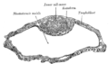 Blastodermic vesicle of Vespertilio murinus.
 -
 Section through embryonic disk of Vespertilio murinus.
 -
 Transverse section of a chorionic villus.
 -
 Scheme of placental circulation.
 -
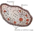
 The initial stages ofhuman embryogenesis
 -
 Histopathology of atubal pregnancy, with labeled cytotrophoblasts and syncytiotrophoblasts.


See also
- Syncytiotrophoblast
- Hydatidiform mole
References
- S2CID 208378249.
- ISBN 978-0-12-815145-7.
- PMID 28110754.
- PMID 19299251.
- PMID 28110755.
- ^ a b "The trophoblast". Archived from the original on 15 December 2007.
- PMID 17288592.
- ^ PMID 31001418.
- ^ PMID 24220516.
External links
- UB, and UF) iperiodembry/carnegie02