
Tunica intima
This article includes a improve this article by introducing more precise citations. (May 2015) ) |
| Tunica intima | |
|---|---|
 | |
 Transverse section through a small artery and vein of the mucous membrane of the epiglottis of a child. (Tunica intima is at "e".) | |
| Details | |
| Part of | Wall of blood vessels |
| Identifiers | |
| Latin | tunica intima |
| MeSH | D017539 |
| TA98 | A12.0.00.018 |
| TA2 | 3922 |
| TH | H3.09.02.0.01003 |
| FMA | 55589 |
| Anatomical terminology | |
The tunica intima (
The three layers of a blood vessel are an inner layer (the tunica intima), a middle layer (the tunica media), and an outer layer (the tunica externa).
In dissection, the inner coat (tunica intima) can be separated from the middle (tunica media) by a little maceration, or it may be stripped off in small pieces; but, because of its friability, it cannot be separated as a complete membrane. It is a fine, transparent, colorless structure which is highly elastic, and, after death, is commonly corrugated into longitudinal wrinkles.
Structure
The structure of the tunica intima depends on the blood vessel type.[1]
Elastic arteries – A single layer of Endothelial and a supporting layer of elastin-rich collagen. The layer also contains fibroblasts and smooth muscle cells called 'myointimal cells'
Arterioles – A single layer of Endothelial cells
Veins – Endothelial cells[2]
The inner coat consists of:
- A layer of pavement endothelium, the cells of which are polygonal, oval, or fusiform, and have very distinct round or oval nuclei. This endothelium is brought into view most distinctly by staining with silver nitrate.
- A subendothelial layer, consisting of delicate connective tissue with branched cells lying in the interspaces of the tissue; in arteries of less than 2 mm in diameter the subendothelial layer consists of a single stratum of stellate cells, and the connective tissue is only largely developed in vessels of a considerable size.[citation needed]
- An elastic or fenestrated layer, which consists of a membrane containing a network of elastic fibers, having principally a longitudinal direction, and in which, under the microscope, small elongated apertures or perforations may be seen, giving it a fenestrated appearance. It was therefore called by Henle the fenestrated membrane. This membrane forms the chief thickness of the inner coat, and can be separated into several layers, some of which present the appearance of a network of longitudinal elastic fibers, and others a more membranous character, marked by pale lines having a longitudinal direction. In minute arteries the fenestrated membrane is a very thin layer; but in the larger arteries, and especially in the aorta, it has a considerable thickness.
Function
Endothelium had been seen to be simply the boundary between the blood in the lumen and the walls of the vessels. However, endothelium has been shown to release local chemicals called
Additional images
-
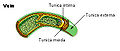 Vein
Vein -
 Microphotography of arterial wall with calcified (violet colour) atherosclerotic plaque (H&E stain)


References
![]() This article incorporates text in the public domain from page 498 of the 20th edition of Gray's Anatomy (1918)
This article incorporates text in the public domain from page 498 of the 20th edition of Gray's Anatomy (1918)
- ^ Steve, Paxton; Michelle, Peckham; Adele, Knibbs (2003). "The Leeds Histology Guide".
{{cite journal}}: Cite journal requires|journal=(help) - ^ Steve, Paxton; Michelle, Peckham; Adele, Knibbs (2003). "The Leeds Histology Guide".
{{cite journal}}: Cite journal requires|journal=(help) - PMID 35625487.
- ISBN 978-1-947172-04-3.
External links
- Histology image: 66_02 at the University of Oklahoma Health Sciences Center – "Aorta"
- Anatomy photo: Circulatory/vessels/vessels7/vessels2 - Comparative Organology at University of California, Davis — "Bird, vessels (LM, High)"
- Image at About.com