
Urethra
| Urethra | |
|---|---|
Deep inguinal lymph nodes | |
| Identifiers | |
| Latin | urethra feminina (female); urethra masculina (male) |
| Greek | οὐρήθρα |
| MeSH | D014521 |
| TA98 | A08.4.01.001F A08.5.01.001M |
| TA2 | 3426, 3442 |
| FMA | 19667 |
| Anatomical terminology] | |
The urethra (pl.: urethras or urethrae) is a tube that connects the mammalian
The external
Structure
The urethra is a fibrous and muscular tube which connects the
Male

In the human male, the urethra is on average 18 to 20 centimeters (7.1 to 7.9 inches) long and opens at the end of the external urethral meatus.[7]
The urethra is divided into four parts in men, named after the location:[7]
| Region | Description | Epithelium |
| pre-prostatic urethra | This is the intramural part of the urethra surrounded by the internal urethral sphincter and varies between 0.5 and 1.5 cm in length depending on the fullness of the bladder. | Transitional |
| prostatic urethra | Crosses through the seminal vesicle, (2) prostatic sinus which has openings for several prostatic ducts where fluid from the prostate enters and contributes to the ejaculate, (3) the prostatic utricle , which is merely an indentation. These openings are collectively called the verumontanum (colliculus seminalis)
The prostatic urethra is a common site of obstruction to outflow of urine in BPH patients |
Transitional |
| membranous urethra | A small (1 or 2 cm) portion passing through the bulbourethral glands (Cowper's gland) are found posterior to this region but open in the spongy urethra . |
Pseudostratified columnar
|
| spongy urethra (or penile urethra) | Runs along the length of the penis on its ventral (underneath) surface. It is about 15 to 25 cm in length, bulbourethral glands are also found here.[9] Some textbooks will subdivide the spongy urethra into two parts, the bulbous and pendulous urethra. The urethral lumen runs effectively parallel to the penis, except at the narrowest point, the external urethral meatus, where it is vertical. This produces a spiral stream of urine and has the effect of cleaning the external urethral meatus. The lack of an equivalent mechanism in the female urethra partly explains why urinary tract infections occur so much more frequently in females. |
Stratified squamous – distally
|
There is inadequate data for the typical length of the male urethra; however, a study of 109 men showed an average length of 22.3 cm (SD = 2.4 cm), ranging from 15 cm to 29 cm.[10]
Female
In the human female, the urethra is about 4 cm long,
Between the
Microanatomy
The cells lining the urethra (the epithelium) start off as transitional cells as it exits the bladder, which are variable layers of flat to cuboidal cells that change shape depending on whether they are compressed by the contents of the urethra.[12] Further along the urethra there are pseudostratified columnar and stratified columnar epithelia.[12] The lining becomes multiple layers of flat cells near the end of the urethra, which is the same as the external skin around it.[12]
There are small mucus-secreting urethral glands, as well as bulbo-urethral glands of Cowper, that secrete mucous acting to lubricate the urethra.[12]
The urethra consists of three coats: muscular, erectile, and mucous, the muscular layer being a continuation of that of the bladder.
Blood and nerve supply and lymphatics
Somatic (conscious) innervation of the external urethral sphincter is supplied by the pudendal nerve.
Development
In the developing
After the third month, urethra also contributes to the development of associated structures depending on the biological sex of the embryo. In the male, the epithelium multiples to form the prostate. In the female, the upper part of the urethra forms the urethra and paraurethral glands.[13]
Function
Urination
The urethra is the vessel through which urine passes after leaving the bladder. During urination, the smooth muscle lining the urethra relaxes in concert with bladder contraction(s) to forcefully expel the urine in a pressurized stream. Following this, the urethra re-establishes muscle tone by contracting the smooth muscle layer, and the bladder returns to a relaxed, quiescent state. Urethral smooth muscle cells are mechanically coupled to each other to coordinate mechanical force and electrical signaling in an organized, unitary fashion.[14]
Ejaculation
The male urethra is the conduit for semen during orgasm.[1] Urine is removed before ejaculation by pre-ejaculate fluid – called Cowper's fluid – from the bulbourethral gland.[15][16]
Clinical significance
Infection of the urethra is
Cancer can also develop in the lining of the urethra.
Injury
Passage of
Injuries to the urethra (e.g., from a pelvic fracture[21])
Foreign bodies in the urethra are uncommon, but there have been medical case reports of self-inflicted injuries, a result of insertion of foreign bodies into the urethra such as an electrical wire.[22]
Other
Hypospadias and epispadias are forms of abnormal development of the urethra in the male, where the meatus is not located at the distal end of the penis (it occurs lower than normal with hypospadias, and higher with epispadias). In a severe chordee, the urethra can develop between the penis and the scrotum.
Catheterisation
A tube called a
Other animals
In all mammals, with the exception of monotremes, and in both sexes, the urethra serves primarily to drain and excrete urine, which in mammals, collects in the urinary bladder and is released from there into the urethra. In addition, the closing mechanisms of the urethra, together with immunoglobulins, largely prevent germs from penetrating the inside of the body.[24] In marsupials, the female's urethra empties into the urogenital sinus.[25]
History
The word "urethra" comes from the
Kidney stones have been identified and recorded about as long as written historical records exist.[27] The urinary tract as well as its function to drain urine from the kidneys, has been described by Galen in the second century AD.[28] Surgery to the urethra to remove kidney stones has been described since at least the first century AD by Aulus Cornelius Celsus.[28]
Additional images
-
 Position of the urethra in males
 -
 Transverse section of the penis
 -
 Male urethral opening on glans penis
 -
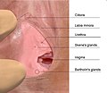 Female urethral opening within vulval vestibule
 -
 Muscles of the female perineum
 -
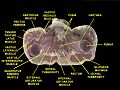 Urethra. Deep dissection. Serial cross section.
 -
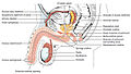 Diagram which depicts the membranous urethra and the spongy urethra of a male
 -


See also
- Perineal urethra
- Vulvovaginal health
- Urethral glands
- Urethral sponge
- Sexual stimulation: Urethral sounding and Urethral intercourse
- Urethrorrhagia
- Urethrotomy
- Urethrectomy
- Internal urethral orifice
References
- ^ ISBN 978-0-226-87013-7. Retrieved 6 May 2013.
- ^ Legato, Marianne J.; Bilezikian, John P., eds. (2004). "109: The Evaluation and Treatment of Urinary Incontinence". Principles of Gender-specific Medicine. Vol. 1. Gulf Professional Publishing. p. 1187.
- PMID 16985861.
- PMID 23094214.
- PMID 15817107.
- from the original on Aug 22, 2023 – via Deep Blue Documents.
- ^ OCLC 920806541.
- ^ "Male Urethra Function & Urethra Anatomy Pictures". Center For Reconstructive Urology. Archived from the original on Sep 26, 2023.
- ^ Atlas of Human Anatomy 5th Edition, Netter.
- PMID 18778496.
- ISBN 9788131225561.
- ^ ISBN 9780702047473.
- ^ ISBN 9781496383907.
- PMID 25483582.
- PMID 21155689.
- S2CID 32553227.
- ^ )
- ^ )
- ^ "Urethral stricture". Mayo Clinic. 12 December 2018. Retrieved 15 May 2020.
- ^ PMID 26816803.
- S2CID 26994715.
- PMID 19192284.
- ^ a b c "Urinary catheters - NHS". nhs.uk. 26 February 2020. Retrieved 2 July 2020.
- ISBN 978-0-521-84158-0.
- ISBN 978-0-521-33792-2.
- ^ S2CID 32778667.
- PMID 24348156.
- ^ PMID 21805756.
External links
- Histology at KUMC epithel-epith07 "Male Urethra"