V(D)J recombination
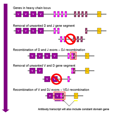
V(D)J recombination (variable–diversity–joining rearrangement) is the mechanism of
V(D)J recombination in mammals occurs in the primary lymphoid organs (
In 1987, Susumu Tonegawa was awarded the Nobel Prize in Physiology or Medicine "for his discovery of the genetic principle for generation of antibody diversity".[1]
Background
Human
- The immunoglobulin heavy locus (IGH@) on chromosome 14, containing the gene segments for the immunoglobulin heavy chain.
- The immunoglobulin kappa (κ) locus (IGK@) on chromosome 2, containing the gene segments for one type (κ) of immunoglobulin light chain.
- The immunoglobulin lambda (λ) locus (IGL@) on chromosome 22, containing the gene segments for another type (λ) of immunoglobulin light chain.
Each heavy chain or light chain gene contains multiple copies of three different types of gene segments for the variable regions of the antibody proteins. For example, the human immunoglobulin heavy chain region contains 2 Constant (Cμ and Cδ) gene segments and 44 Variable (V) gene segments, plus 27 Diversity (D) gene segments and 6 Joining (J) gene segments.[2] The light chain genes possess either a single (Cκ) or four (Cλ) Constant gene segments with numerous V and J gene segments but do not have D gene segments.[3] DNA rearrangement causes one copy of each type of gene segment to go in any given lymphocyte, generating an enormous antibody repertoire; roughly 3×10¹¹ combinations are possible, although some are removed due to self reactivity.
Most T cell receptors are composed of a variable alpha chain and a beta chain. The T cell receptor genes are similar to immunoglobulin genes in that they too contain multiple V, D, and J gene segments in their beta chains (and V and J gene segments in their alpha chains) that are rearranged during the development of the lymphocyte to provide that cell with a unique antigen receptor. The T cell receptor in this sense is the topological equivalent to an antigen-binding fragment of the antibody, both being part of the immunoglobulin superfamily.
An autoimmune response is prevented by eliminating cells that self-react. This occurs in the thymus by testing the cell against an array of self antigens expressed through the function of the autoimmune regulator (AIRE). The immunoglobulin lambda light chain locus contains protein-coding genes that can be lost with its rearrangement. This is based on a physiological mechanism and is not pathogenetic for leukemias or lymphomas. A cell persists if it creates a successful product that does not self-react, otherwise it is pruned via apoptosis.
Immunoglobulins

Heavy chain
In the developing
Light chain
The kappa (κ) and lambda (λ) chains of the immunoglobulin light chain loci rearrange in a very similar way, except that the light chains lack a D segment. In other words, the first step of recombination for the light chains involves the joining of the V and J chains to give a VJ complex before the addition of the constant chain gene during primary transcription. Translation of the spliced mRNA for either the kappa or lambda chains results in formation of the Ig κ or Ig λ light chain protein.
Assembly of the Ig μ heavy chain and one of the light chains results in the formation of membrane bound form of the immunoglobulin IgM that is expressed on the surface of the immature B cell.
T cell receptors
During
The rearrangement of the alpha (α) chain of the TCR follows β chain rearrangement, and resembles V-to-J rearrangement described for Ig light chains (see above). The assembly of the β- and α- chains results in formation of the αβ-TCR that is expressed on a majority of T cells.
Mechanism
Key enzymes and components
The process of V(D)J recombination is mediated by VDJ recombinase, which is a diverse collection of enzymes. The key enzymes involved are recombination activating genes 1 and 2 (RAG), terminal deoxynucleotidyl transferase (TdT), and Artemis nuclease, a member of the ubiquitous non-homologous end joining (NHEJ) pathway for DNA repair.[4] Several other enzymes are known to be involved in the process and include DNA-dependent protein kinase (DNA-PK), X-ray repair cross-complementing protein 4 (XRCC4), DNA ligase IV, non-homologous end-joining factor 1 (NHEJ1; also known as Cernunnos or XRCC4-like factor [XLF]), the recently discovered Paralog of XRCC4 and XLF (PAXX), and DNA polymerases λ and μ.[5] Some enzymes involved are specific to lymphocytes (e.g., RAG, TdT), while others are found in other cell types and even ubiquitously (e.g., NHEJ components).
To maintain the specificity of recombination, V(D)J recombinase recognizes and binds to
Process
V(D)J recombination begins when V(D)J recombinase (through the activity of RAG1) binds a RSS flanking a coding gene segment (V, D, or J) and creates a single-strand nick in the DNA between the first base of the RSS (just before the heptamer) and the coding segment. This is essentially energetically neutral (no need for
The blunt signal ends are flush ligated together to form a circular piece of DNA containing all of the intervening sequences between the coding segments known as a signal joint (although circular in nature, this is not to be confused with a plasmid). While originally thought to be lost during successive cell divisions, there is evidence that signal joints may re-enter the genome and lead to pathologies by activating oncogenes or interrupting tumor suppressor gene function(s)[Ref].
The coding ends are processed further prior to their ligation by several events that ultimately lead to junctional diversity.[15] Processing begins when DNA-PK binds to each broken DNA end and recruits several other proteins including Artemis, XRCC4, DNA ligase IV, Cernunnos, and several DNA polymerases.[16] DNA-PK forms a complex that leads to its autophosphorylation, resulting in activation of Artemis. The coding end hairpins are opened by the activity of Artemis.[17] If they are opened at the center, a blunt DNA end will result; however in many cases, the opening is "off-center" and results in extra bases remaining on one strand (an overhang). These are known as palindromic (P) nucleotides due to the palindromic nature of the sequence produced when DNA repair enzymes resolve the overhang.[18] The process of hairpin opening by Artemis is a crucial step of V(D)J recombination and is defective in the severe combined immunodeficiency (scid) mouse model.
Next, XRCC4, Cernunnos, and DNA-PK align the DNA ends and recruit terminal deoxynucleotidyl transferase (TdT), a template-independent DNA polymerase that adds non-templated (N) nucleotides to the coding end. The addition is mostly random, but TdT does exhibit a preference for G/C nucleotides.[19] As with all known DNA polymerases, the TdT adds nucleotides to one strand in a 5' to 3' direction.[20]
Lastly, exonucleases can remove bases from the coding ends (including any P or N nucleotides that may have formed). DNA polymerases λ and μ then insert additional nucleotides as needed to make the two ends compatible for joining. This is a stochastic process, therefore any combination of the addition of P and N nucleotides and exonucleolytic removal can occur (or none at all). Finally, the processed coding ends are ligated together by DNA ligase IV.[21]
All of these processing events result in a
See also
- B cell receptor
- T cell receptor
- Basel Institute for Immunology
- Charles M. Steinberg
- NKT cell
- Recombination-activating gene
References
- ^ "The Nobel Prize in Physiology or Medicine 1987". nobelprize.org. Archived from the original on 13 February 2021. Retrieved 26 December 2014.
- PMID 15010366.
- ^ ISBN 978-0-323-47978-3.
- PMID 16082219.
- S2CID 45771818.
- PMID 8208601.
- S2CID 40771963.
- PMID 8620529.
- PMID 9651584.
- PMID 21854230.
- S2CID 33489235.
- S2CID 10975251.
- PMID 9094713.
- PMID 10837067.
- PMID 8073949.
- PMID 22224778.
- PMID 15936993.
- PMID 17932067.
- PMID 8524303.
- S2CID 20634390.
- PMID 18066085.
Further reading
- Hartwell LH, Hood L, Goldberg ML, Reynolds AE, Silver LM, Veres RC (2000). Chapter 24, Evolution at the molecular level. In: Genetics. New York: McGraw-Hill. pp. 805–807. ISBN 978-0-07-299587-9.
- V(D)J Recombination. Series: Advances in Experimental Medicine and Biology, Vol. 650 Ferrier, Pierre (Ed.) Landes Bioscience 2009, XII, 199 p. ISBN 978-1-4419-0295-5
