
Vertebra
| Vertebra | |
|---|---|
Spinal column | |
| Identifiers | |
| Latin | vertebra |
| TA98 | A02.2.01.001 |
| TA2 | 1011 |
| FMA | 9914 |
| Anatomical terms of bone] | |
Each vertebra (pl.: vertebrae) is an irregular bone with a complex structure composed of bone and some hyaline cartilage, that make up the vertebral column or spine, of vertebrates. The proportions of the vertebrae differ according to their spinal segment and the particular species.
The basic configuration of a vertebra varies; the bone is the body, and the central part of the body is the centrum. The upper and lower surfaces of the vertebra body give attachment to the
Vertebrae articulate with each other to give strength and flexibility to the spinal column, and the shape at their back and front aspects determines the range of movement. Structurally, vertebrae are essentially alike across the vertebrate species, with the greatest difference seen between an aquatic animal and other vertebrate animals. As such, vertebrates take their name from the vertebrae that compose the vertebral column.
Structure
In the human vertebral column, the size of the vertebrae varies according to placement in the vertebral column, spinal loading, posture and pathology. Along the length of the spine, the vertebrae change to accommodate different needs related to stress and mobility.[1] Each vertebra is an irregular bone.

A typical vertebra has a body (vertebral body), which consists of a large anterior middle portion called the centrum (vertebral centrum, plural centra) and a posterior vertebral arch,

The vertebral arch is formed by pedicles and laminae. Two pedicles extend from the sides of the vertebral body to join the body to the arch. The pedicles are short thick processes that extend, one from each side, posteriorly, from the junctions of the posteriolateral surfaces of the centrum, on its upper surface.
From each pedicle a broad plate, a lamina, projects backward and medially to join and complete the vertebral arch and form the posterior border of the vertebral foramen, which completes the triangle of the vertebral foramen.
Processes
There are seven processes projecting from the vertebra:
- one spinous process
- two transverse processes
- four articular processes
A major part of a vertebra is a backward extending spinous process (sometimes called the neural spine) which projects centrally..
The two transverse processes, one on each side of the vertebral body, project laterally from either side at the point where the lamina joins the
There are superior and inferior articular facet joints on each side of the vertebra, which serve to restrict the range of movement possible. These facets are joined by a thin portion of the vertebral arch called the pars interarticularis.
Regional variation

Vertebrae take their names from the regions of the vertebral column that they occupy. There are usually thirty-three vertebrae in the human vertebral column — seven
The regional vertebrae increase in size as they progress downward but become smaller in the coccyx.
Cervical

There are seven
Cervical vertebrae possess transverse foramina to allow for the vertebral arteries to pass through on their way to the foramen magnum to end in the circle of Willis. These are the smallest, lightest vertebrae and the vertebral foramina are triangular in shape. The spinous processes are short and often bifurcated (the spinous process of C7 is not bifurcated, and is substantially longer than that of the other cervical spinous processes).[16]
The atlas differs from the other vertebrae in that it has no body and no spinous process. It has instead a ring-like form, having an anterior and a posterior arch and two lateral masses. At the outside centre points of both arches there is a tubercle, an anterior tubercle and a posterior tubercle, for the attachment of muscles. The front surface of the anterior arch is convex and its anterior tubercle gives attachment to the
Specific to the cervical vertebra is the transverse foramen (also known as foramen transversarium). This is an opening on each of the transverse processes which gives passage to the vertebral artery and vein and a sympathetic nerve plexus. On the cervical vertebrae other than the atlas, the anterior and posterior tubercles are on either side of the transverse foramen on each transverse process. The anterior tubercle on the sixth cervical vertebra is called the carotid tubercle because it separates the carotid artery from the vertebral artery.
There is a hook-shaped uncinate process on the side edges of the top surface of the bodies of the third to the seventh cervical vertebrae and of the first thoracic vertebra. Together with the vertebral disc, this uncinate process prevents a vertebra from sliding backward off the vertebra below it and limits lateral flexion (side-bending). Luschka's joints involve the vertebral uncinate processes.
The spinous process on C7 is distinctively long and gives the name
The term cervicothoracic is often used to refer to the cervical and thoracic vertebrae together, and sometimes also their surrounding areas.
Thoracic

The twelve thoracic vertebrae and their transverse processes have surfaces that articulate with the ribs. Some rotation can occur between the thoracic vertebrae, but their connection with the rib cage prevents much flexion or other movement. They may also be known as "dorsal vertebrae" in the human context.
The vertebral bodies are roughly heart-shaped and are about as wide anterio-posteriorly as they are in the transverse dimension. Vertebral foramina are roughly circular in shape.
The top surface of the first thoracic vertebra has a hook-shaped uncinate process, just like the cervical vertebrae.
The thoracolumbar division refers to the thoracic and lumbar vertebrae together, and sometimes also their surrounding areas.
The thoracic vertebrae attach to ribs and so have articular facets specific to them; these are the superior, transverse and inferior costal facets. As the vertebrae progress down the spine they increase in size to match up with the adjoining lumbar section.
Lumbar


The five lumbar vertebrae are the largest of the vertebrae, their robust construction being necessary for supporting greater weight than the other vertebrae. They allow significant flexion, extension and moderate lateral flexion (side-bending). The discs between these vertebrae create a natural lumbar lordosis (a spinal curvature that is concave posteriorly).[citation needed] This is due to the difference in thickness between the front and back parts of the intervertebral discs.
The lumbar vertebrae are located between the ribcage and the pelvis and are the largest of the vertebrae. The pedicles are strong, as are the laminae, and the spinous process is thick and broad. The vertebral foramen is large and triangular. The transverse processes are long and narrow and three tubercles can be seen on them. These are a lateral costiform process, a mammillary process and an accessory process.[17] The superior, or upper tubercle is the mammillary process which connects with the superior articular process. The multifidus muscle attaches to the mammillary process and this muscle extends through the length of the vertebral column, giving support. The inferior, or lower tubercle is the accessory process and this is found at the back part of the base of the transverse process. The term lumbosacral is often used to refer to the lumbar and sacral vertebrae together, and sometimes includes their surrounding areas.
Sacral

There are five sacral vertebrae (S1–S5) which are fused in maturity, into one large bone, the
Coccygeal
The last three to five coccygeal vertebrae (but usually four) (Co1–Co5) make up the tailbone or coccyx.[19] There are no intervertebral discs.
Development

Function
Functions of vertebrae include:
- Support of the vertebrae function in the skeletomuscular system by forming the vertebral column to support the body
- Protection. Vertebrae contain a vertebral foramen for the passage of the spinal canal and its enclosed spinal cord and covering meninges. They also afford sturdy protection for the spinal cord. The upper and lower surfaces of the centrum are flattened and rough in order to give attachment to the intervertebral discs.
- Movement. The vertebrae also provide the openings, the intervertebral foramina which allow the entry and exit of the spinal nerves. Similarly to the surfaces of the centrum, the upper and lower surfaces of the fronts of the laminae are flattened and rough to give attachment to the ligamenta flava. Working together in the vertebral column their sections provide controlled movement and flexibility.
- Feeding of the intervertebral discs through the reflex (hyaline ligament) plate that separates the cancellous bone of the vertebral body from each disk
-
 The spinal cord nested in the vertebral column.
 -
 Vertebral joint
 -
 Costovertebral joint
 -
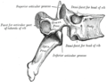 A facet joint between the superior and inferior articular processes (labeled at top and bottom).


Clinical significance
There are a number of
Spondylolysis is a defect in the pars interarticularis of the vertebral arch. In most cases this occurs in the lowest of the lumbar vertebrae (L5), but may also occur in the other lumbar vertebrae, as well as in the thoracic vertebrae.
A laminectomy is a surgical operation to remove the laminae in order to access the spinal canal.[21] The removal of just part of a lamina is called a laminotomy.
A
Another condition is spondylolisthesis when one vertebra slips forward onto another. The reverse of this condition is retrolisthesis where one vertebra slips backward onto another.
The vertebral pedicle is often used as a radiographic marker and entry point in
The arcuate foramen is a common anatomical variation more frequently seen in females. It is a bony bridge found on the first cervical vertebra, the atlas where it covers the groove for the vertebral artery.[22]
Degenerative disc disease is a condition usually associated with ageing in which one or more discs degenerate. This can often be a painfree condition but can also be very painful.
Non-humans

In other animals, the vertebrae take the same regional names except for the coccygeal – in animals with tails, the separate vertebrae are usually called the caudal vertebrae.
Vertebrae with saddle-shaped articular surfaces on their bodies, called "heterocoelous", allow vertebrae to flex both vertically and horizontally while preventing twisting motions. Such vertebrae are found in the necks of birds and some turtles.[23]

"Procoelous" vertebrae feature a spherical protrusion extending from the caudal end of the centrum of one vertebra that fits into a concave socket on the cranial end of the centrum of an adjacent vertebra.
In many species, though not in mammals, the cervical vertebrae bear ribs. In many groups, such as
In all mammals, the thoracic vertebrae are connected to
There are fewer lumbar vertebrae in chimpanzees and gorillas, which have three in contrast to the five in the genus Homo. This reduction in number gives an inability of the lumbar spine to lordose but gives an anatomy that favours vertical climbing, and hanging ability more suited to feeding locations in high-canopied regions.[34] The bonobo differs by having four lumbar vertebrae.
Additional images
-
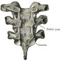 Vertebral arches of three thoracic vertebrae
 -
 Costovertebral joints seen from the front
 -
 Lower thoracic and upper lumbar vertebrae seen from the side
 -
 Cervical vertebrae seen from the back
 -
Vertebrae anatomy


See also
- Limbus vertebra
- Functional spinal unit
- Pott disease
- Scheuermann's disease
References
![]() This article incorporates text in the public domain from page 96 of the 20th edition of Gray's Anatomy (1918)
This article incorporates text in the public domain from page 96 of the 20th edition of Gray's Anatomy (1918)
- ^ McGraw-Hill Science and Technology[full citation needed]
- ^ O'Rahilly, Müller, Carpenter & Swenson. "Chapter 39: The vertebral column". Basic Human Anatomy. www.dartmouth.edu.
{{cite web}}: CS1 maint: multiple names: authors list (link) - ISBN 978-1-4160-6257-8.
- S2CID 206078730.
- PMID 18193299.
- ^ Taylor, Tim. "Lumbar Vertebrae". InnerBody. Retrieved May 7, 2017.
- ^ ISBN 978-0-323-07954-9, retrieved 2020-11-03
- ^ Standring, Susan (2008). "Thoracic vertebrae". Gray's Anatomy. p. 746.
- ^ Platzer (2004), pp 42–43
- ^ Latin costa refers to either a "rib" or "side" of the body. (Diab (1999), p 76)
- ^ a b Tweedie, A. The library of medicine p. 31
- ^ Heinz Feneis, Wolfgang Dauber (2000) Pocket Atlas of Human Anatomy: Based on the International Nomenclature p. 2
- ISBN 978-0-12-374247-6. Retrieved 1 November 2023.
- PMID 27559975. Retrieved 1 November 2023.
- ^ PMID 34935276.
- ISBN 978-0-323-07954-9, retrieved 2020-11-03
- ^ Postacchini, Franco (1999) Lumbar Disc Herniation p. 19
- ^ Drake et al, Gray's Anatomy for Students, Churchill Livingstone/Elsevier (2010), 2nd edition, chapter 2
- ^ a b Weisberger, Mindy (March 23, 2024). "Why don't humans have tails? Scientists find answers in an unlikely place". CNN. Archived from the original on March 24, 2024. Retrieved March 24, 2024.
- ^ a b Walker, Warren F., Jr. (1987) Functional Anatomy of the Vertebrate San Francisco: Saunders College Publishing.
- ISBN 978-1-4160-6257-8.
- PMID 16155658.
- ISBN 978-0-07-290956-2.
- ^ Romer, Alfred (1962). The Vertebrate Body (3 ed.). Philadelphia, Pa; London, W.C.I.: W.B. Saunders Company.
- ^ PMC 2263429.
- ^ Romer, Alfred (1956). Osteology of the Reptiles. Malabar, Florida: Krieger Publishing Company. pp. 0–89464–985–X.
- ^ ISBN 978-0-07-802302-6.
- ^ "Beluga Whale". Yellowmagpie.com. 2012-06-27. Retrieved 2012-08-12.
- ^ "About Whales". Whalesalive.org.au. 2009-06-26. Retrieved 2013-08-12.
- ISBN 9781420041637. Retrieved May 7, 2017.
- ^ Hyman, Libbie (1922). Comparative Vertebrate Anatomy. Chicago: University of Chicago Press. pp. 123.
- ^ "Physical Characteristics of the Koala". Australian Koala Foundation. Retrieved 1 February 2012.
- ^ Hyman (1922), p. 124
- PMID 20855303.)
{{cite journal}}: CS1 maint: multiple names: authors list (link - ISBN 978-0-07-290956-2.
- ^ Hyman, Libbie (1922). Comparative Vertebrate Anatomy. Chicago: University of Chicago Press. pp. 125.
External links
- Atlas image: back_bone13 at the University of Michigan Health System – Axis & Atlas Articulated, Posterior View
- Anatomy image: skel/atlas2 at Human Anatomy Lecture (Biology 129), Pennsylvania State University
- Anatomy photo:26:os-0110 at the SUNY Downstate Medical Center
- Anatomy figure: 02:01-10 at Human Anatomy Online, SUNY Downstate Medical Center
- Anatomy figure: 18:02-01 at Human Anatomy Online, SUNY Downstate Medical Center
- Vertebra - BlueLink Anatomy - University of Michigan Medical School
- Atlas image: back_bone28 at the University of Michigan Health System
- Anatomy figure: 02:02-08 at Human Anatomy Online, SUNY Downstate Medical Center
- The shapes of the articulating ends of vertebrae – University of the Cumberlands