
Vocal cords
| Vocal cords | |
|---|---|
adduction | |
| Details | |
| Precursor | Sixth pharyngeal arch |
| System | Respiratory system |
| Identifiers | |
| Latin | plica vocalis |
| MeSH | D014827 |
| TA98 | A06.2.09.013 |
| TA2 | 3198 |
| FMA | 55457 |
| Anatomical terminology] | |


In humans, the vocal cords, also known as vocal folds, are folds of throat tissues that are key in creating sounds through vocalization. The size of vocal cords affects the pitch of voice. Open when breathing and vibrating for speech or singing, the folds are controlled via the recurrent laryngeal branch of the vagus nerve. They are composed of twin infoldings of mucous membrane stretched horizontally, from back to front, across the larynx. They vibrate, modulating the flow of air being expelled from the lungs during phonation.[1]
The 'true vocal cords' are distinguished from the 'false vocal folds', known as vestibular folds or ventricular folds, which sit slightly superior to the more delicate true folds. These have a minimal role in normal phonation, but can produce deep sonorous tones, screams and growls.
The length of the vocal fold at birth is approximately six to eight millimeters and grows to its adult length of eight to sixteen millimeters by adolescence. DHT, an androgen metabolite of testosterone which is secreted by the gonads, causes changes in the cartilages and musculature of the larynx when present in high enough concentrations, such as during an adolescent boy's puberty: The thyroid prominence appears, the vocal folds lengthen and become rounded, and the epithelium thickens with the formation of three distinct layers in the lamina propria.[citation needed]. These changes are only partially reversible via reconstructive surgery such as Chondrolaryngoplasty, Feminization laryngoplasty, and laser tuning of the vocal cords.
Structure
Location
The vocal folds are located within the larynx at the top of the
False vocal folds
The vocal folds are sometimes called 'true vocal folds' to distinguish them from the 'false vocal folds' known as
Microanatomy
The vocal cords are composed of twin infoldings of 3 distinct tissues: an outer layer of flat cells that do not produce
Variation
Males and females have different vocal fold sizes. Adult male
Development
In newborns
Newborns have a uniform single layered lamina propria, which appears loose with no vocal ligament.[4] The monolayered lamina propria is composed of ground substances such as hyaluronic acid and fibronectin, fibroblasts, elastic fibers, and collagenous fibers. While the fibrous components are sparse, making the lamina propria structure loose, the hyaluronic acid (HA) content is high.
HA is a bulky, negatively charged glycosaminoglycan, whose strong affinity with water procures hyaluronic acid its viscoelastic and shock absorbing properties essential to vocal biomechanics.[5] Viscosity and elasticity are critical to voice production. Chan, Gray and Titze, quantified the effect of hyaluronic acid on both the viscosity and the elasticity of vocal folds by comparing the properties of tissues with and without HA.[6] The results showed that removal of hyaluronic acid decreased the stiffness of the vocal cords by an average of 35%, but increased their dynamic viscosity by an average of 70% at frequencies higher than 1 Hz. Newborns have been shown to cry an average of 6.7 hours per day during the first 3 months, with a sustained pitch of 400–600 Hz, and a mean duration per day of 2 hours.[7] Similar treatment on adult vocal cords would quickly result in edema, and subsequently aphonia. Schweinfurth and al. presented the hypothesis that high hyaluronic acid content and distribution in newborn vocal cords is directly associated with newborn crying endurance.[7] These differences in newborn vocal fold composition would also be responsible for newborns inability to articulate sounds, besides the fact that their lamina propria is a uniform structure with no vocal ligament. The layered structure necessary for phonation will start to develop during the infancy and until the adolescence.[4]
The fibroblasts in the newborn Reinke's space are immature, showing an oval shape, and a large nucleus-cytoplasm ratio.[4] The rough endoplasmic reticulum and Golgi apparatus, as shown by electron micrographs, are not well developed, indicating that the cells are in a resting phase. The collagenous and reticular fibers in the newborn the vocal cords are fewer than in the adult one, adding to the immaturity of the vocal fold tissue.
In the infant, many fibrous components were seen to extend from the macula flava towards the Reinke's space. Fibronectin is very abundant in the Reinke's space of newborn and infant. Fibronectin is a glycoprotein that is believed to act as a template for the oriented deposition of the collagen fibers, stabilizing the collagen fibrils. Fibronectin also acts as a skeleton for the elastic tissue formation.[4] Reticular and collagenous fibers were seen to run along the edges of the vocal cords throughout the entire lamina propria.[4] Fibronectin in the Reinke's space appeared to guide those fibers and orient the fibril deposition. The elastic fibers remained sparse and immature during infancy, mostly made of microfibrils. The fibroblasts in the infant Reinke's space were still sparse but spindle-shaped. Their rough endoplasmic reticulum and Golgi apparatus were still not well developed, indicating that despite the change in shape, the fibroblasts still remained mostly in a resting phase. Few newly released materials were seen adjacent to the fibroblasts. The ground substance content in the infant Reinke's space seemed to decrease over time, as the fibrous component content increased, thus slowly changing the vocal fold structure.
Children
The infant lamina propria is composed of only one layer, as compared to three in the adult, and there is no vocal ligament. The vocal ligament begins to be present in children at about four years of age. Two layers appear in the lamina propria between the ages of six and twelve, and the mature lamina propria, with the superficial, intermediate and deep layers, is only present by the conclusion of adolescence. As vocal fold vibration is a foundation for vocal formants, this presence or absence of tissue layers influences a difference in the number of formants between the adult and pediatric populations. In females, the voice is three tones lower than the child's and has five to twelve formants, as opposed to the pediatric voice with three to six. The length of the vocal fold at birth is approximately six to eight millimeters and grows to its adult length of eight to sixteen millimeters by adolescence. The infant vocal fold is half membranous or anterior glottis, and half cartilaginous or posterior glottis. The adult fold is approximately three-fifths membranous and two-fifths cartilaginous.
Puberty
Adulthood
Human vocal cords are paired structures located in the larynx, just above the trachea, which vibrate and are brought in contact during phonation. The human vocal cords are roughly 12 – 24 mm in length, and 3–5 mm thick.[9] Histologically, the human vocal cords are a laminated structure composed of five different layers. The vocalis muscle, main body of the vocal cords, is covered by the mucosa, which consists of the epithelium and the lamina propria.[10] The latter is a pliable layer of connective tissue subdivided into three layers: the superficial layer (SL), the intermediate layer (IL), and the deep layer (DL).[11] Layer distinction is either made looking at differential in cell content or extracellular matrix (extracellular matrix) content. The most common way being to look at the extracellular matrix content. The SLP has fewer elastic and collagenous fibers than the two other layers, and thus is looser and more pliable. The ILP is mostly composed of elastic fibers, while the DLP has fewer elastic fibers, and more collagenous fibers.[10] In those two layers, which form what is known as the vocalis ligament, the elastic and collagenous fibers are densely packed as bundles that run almost parallel to the edge of the vocal fold.[10]
There is a steady increase in the elastin content of the lamina propria as humans age (elastin is a yellow scleroprotein, the essential constituent of the elastic connective tissue) resulting in a decrease in the ability of the lamina propria to expand caused by cross-branching of the elastin fibers. Among other things, this leads to the mature voice being better suited to the rigors of opera.[citation needed]
The extracellular matrix of the vocal cord LP is composed of fibrous proteins such as collagen and elastin, and interstitial molecules such as
Maturation
Vocal fold structure in adults is quite different from that in newborns. Exactly how the vocal cord mature from an immature monolayer in newborns to a mature three layer tissue in adults is still unknown, however a few studies have investigated the subjects and brought some answers.
Hirano et al. previously found that the newborns did not have a true lamina propria, but instead had cellular regions called maculae flavae, located at the anterior and posterior ends of the loose vocal fold tissue.[4][14] Boseley and Hartnick examined at the development and maturation of pediatric human vocal fold lamina propria.[15] Hartnick was the first one to define each layer by a change in their cellular concentration.[16] He also found that the lamina propria monolayer at birth and shortly thereafter was hypercellular, thus confirming Hirano's observations. By 2 months of age, the vocal fold started differentiating into a bilaminar structure of distinct cellular concentration, with the superficial layer being less densely populated than the deeper layer. By 11 months, a three-layered structure starts to be noted in some specimens, again with different cellular population densities. The superficial layer is still hypocellular, followed by an intermediate more hypercellular layer, and a deeper hypercellular layer, just above the vocalis muscle. Even though the vocal cords seem to start organizing, this is not representative of the trilaminar structure seen in adult tissues, where the layer are defined by their differential elastin and collagen fiber compositions. By 7 years of age, all specimens show a three-layered vocal fold structure, based on cellular population densities. At this point, the superficial layer was still hypocellular, the middle layer was the hypercellular one, with also a greater content of elastin and collagen fibers, and the deeper layer was less cellularly populated. Again, the distinction seen between the layers at this stage is not comparable to that seen in the adult tissue. The maturation of the vocal cords did not appear before 13 years of age, where the layers could be defined by their differential fiber composition rather than by their differential cellular population. The pattern now show a hypocellular superficial layer, followed by a middle layer composed predominantly of elastin fiber, and a deeper layer composed predominantly of collagen fibers. This pattern can be seen in older specimens up to 17 years of age, and above. While this study offers a nice way to see the evolution from immature to mature vocal cords, it still does not explain what is the mechanism behind it.
Macula flavae
Maculae flavae are located at the anterior and posterior ends of the membranous parts of the vocal cords.
Impact of phonation
The viscoelastic properties of human vocal fold lamina propria are essential for their vibration, and depend on the composition and structure of their extracellular matrix. Adult vocal cords have a layered structure which is based on the layers differential in extracellular matrix distribution. Newborns on the other hand, do not have this layered structure. Their vocal cords are uniform, and immature, making their viscoelastic properties most likely unsuitable for phonation. Hyaluronic acid plays a very important role in the vocal fold biomechanics. In fact, hyaluronic acid has been described as the extracellular matrix molecule that not only contributes to the maintenance of an optimal tissue viscosity that allows phonation, but also of an optimal tissue stiffness that allows frequency control.[6] CD44 is a cell surface receptor for HA. Cells such as fibroblasts are responsible for synthesizing extracellular matrix molecules. Cell surface matrix receptors in return, feed back to the cells through cell-matrix interaction, allowing the cell to regulate its metabolism.
Sato et al.[18] carried out a histopathologic investigation of unphonated human vocal cords. Vocal fold mucosae, which were unphonated since birth, of three young adults (17, 24, and 28 years old) were looked at using light and electron microscopy. The results show that the vocal fold mucosae were hypoplastic, and rudimentary, and like newborns, did not have any vocal ligament, Reinke's space, or layered structure. Like newborns, the lamina propria appeared as a uniform structure. Some stellate cells were present in the macula flava, but started to show some signs of degeneration. The stellate cells synthesized fewer extracellular matrix molecules, and the cytoplasmic processes were shown to be short and shrinking, suggesting a decreased activity. Those results confirm the hypothesis that phonation stimulates stellate cells into producing more extracellular matrix.
Furthermore, using a specially designed bioreactor, Titze et al. showed that fibroblasts exposed to mechanical stimulation have differing levels of extracellular matrix production from fibroblasts that are not exposed to mechanical stimulation.[19] The gene expression levels of extracellular matrix constituents such as fibronectin, MMP1, decorin, fibromodulin, hyaluronic acid synthase 2, and CD44 were altered. All those genes are involved in extracellular matrix remodeling, thus suggesting that mechanical forces applied to the tissue, alter the expression levels of extracellular matrix related genes, which in turn allow the cells present in the tissue to regulate the extracellular matrix constituent synthesis, thus affecting the tissue's composition, structure, and biomechanical properties. In the end, cell-surface receptors close the loop by giving feedback on the surrounding extracellular matrix to the cells, affecting also their gene expression level.
Impact of hormones
Other studies suggest that
Vocal fold phonatory functions are known to change from birth to old age. The most significant changes occur in development between birth and puberty, and in old age.
Old age
There is a thinning in the superficial layer of the lamina propria in old age. In aging, the vocal fold undergoes considerable sex-specific changes. In the female larynx, the vocal fold cover thickens with aging. The superficial layer of the lamina propria loses density as it becomes more edematous. The intermediate layer of the lamina propria tends to atrophy only in men. The deep layer of the lamina propria of the male vocal fold thickens because of increased collagen deposits. The vocalis muscle atrophies in both men and women. However, the majority of elderly patients with voice disorders have disease processes associated with aging rather than physiologic aging alone.[25][26][27]
Function
Oscillation

The larynx is a major (but not the only) source of sound in speech, generating sound through the rhythmic opening and closing of the vocal folds. To oscillate, the vocal folds are brought near enough together such that air pressure builds up beneath the larynx. The folds are pushed apart by this increased subglottal pressure, with the inferior part of each fold leading the superior part. Such a wave-like motion causes a transfer of energy from the airflow to the fold tissues.[28] Under the correct conditions, the energy transferred to the tissues is large enough to overcome losses by dissipation and the oscillation pattern will sustain itself. In essence, sound is generated in the larynx by chopping up a steady flow of air into little puffs of sound waves.[29]
The perceived pitch of a person's voice is determined by a number of different factors, most importantly the
The vocal folds generate a sound rich in harmonics. The harmonics are produced by collisions of the vocal folds with themselves, by recirculation of some of the air back through the trachea, or both.[31] Some singers can isolate some of those harmonics in a way that is perceived as singing in more than one pitch at the same time—a technique called overtone singing or throat singing such as in the tradition of Tuvan throat singing.
Clinical significance
Lesions
The majority of vocal fold lesions primarily arise in the cover of the folds. Since the basal lamina secures the epithelium to the superficial layer of the lamina propria with anchoring fibers, this is a common site for injury. If a person has a phonotrauma or habitual vocal hyperfunction, also known as pressed phonation, the proteins in the basal lamina can shear, causing vocal fold injury, usually seen as nodules or polyps, which increase the mass and thickness of the cover. The
Reinke's edema
A voice pathology called Reinke's edema, swelling due to abnormal accumulation of fluid, occurs in the superficial lamina propria or Reinke's space. This causes the vocal fold mucosa to appear floppy with excessive movement of the cover that has been described as looking like a loose sock.[32] The greater mass of the vocal folds due to increased fluid lowers the fundamental frequency during phonation.
Wound healing
Wound healing is a natural regeneration process of dermal and epidermal tissue involving a sequence of biochemical events. These events are complex and can be categorized into three stages: inflammation, proliferation and tissue remodeling.
Any injury to human vocal folds elicits a wound healing process characterized by disorganized collagen deposition and, eventually, formation of scar tissue.
Terminology
The vocal folds are commonly referred to as vocal cords, and less commonly as vocal flaps or vocal bands. The term vocal cords was coined by the French anatomist Antoine Ferrein in 1741. In his violin analogy of the human voice, he postulated that the moving air acted like a bow on cordes vocales.[42] The alternative spelling in English is vocal chords, possibly due to the musical connotations or to confusion with the geometrical definition of the word chord. While both spellings have historical precedents, standard American spelling is cords.[43] According to the Oxford English Corpus, a database of 21st-century texts that contains everything from academic journal articles to unedited writing and blog entries, contemporary writers opt for the nonstandard chords instead of cords 49% of the time.[44][45] The cords spelling is also standard in the United Kingdom and Australia.
In phonetics, vocal folds is preferred over vocal cords, on the grounds that it is more accurate and illustrative.[46][47][48]
See also
- Adam's apple
- Electroglottograph
- Vocal register
- Falsetto
- Vocal fry
- Vocal cord dysfunction
- Vocology
- Articulatory phonetics
- Laryngospasm
Additional images
-
 Vocal folds.
 -
 Coronal section of larynx and upper part of trachea.
 -
 The entrance to the larynx, viewed from behind.
 -
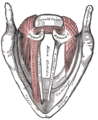 Muscles of the larynx, seen from above.


References
- S2CID 33929329.
- ^ Fuks, Leonardo (1998). "From Air to Music: Acoustical, Physiological and Perceptual Aspects of Reed Wind Instrument Playing and Vocal-Ventricular Fold Phonation". Stockholm, Sweden. Archived from the original on 2009-12-27. Retrieved 2010-01-05.
- ISBN 978-0-13-717893-3. Archivedfrom the original on 2017-09-05.
- ^ S2CID 22257136.
- ^ PMID 12395982.
- ^ PMID 11391249.
- ^ S2CID 35188777.
- ^ Abitbol, A. & Abitbol, P. (2003). The Larynx: A Hormonal Target. In Rubin, J.S., Sataloff, R.T., & Korovin, G.S. (Eds.), Diagnosis and Treatment of Voice Disorders (pp. 355-380). Clifton Park, NY: Delmar Learning.
- PMID 16154633.
- ^ a b c d e Hirano, M., S. Kurita, and T. Nakashima. Vocal fold physiology : contemporary research and clinical issues. in Vocal Fold Physiology, Conference. 1981. San Diego, Calif.: College-Hill Press.
- ^ PMID 10918654.
- ^ PMID 9075177.
- S2CID 71920488.
- S2CID 32824702.
- S2CID 21613826.
- S2CID 6024918.
- S2CID 12529469.
- S2CID 21410937.
- PMID 15336927.
- ^ PMID 18852972.
- ^ PMID 10764118.
- PMID 2756834.
- ISBN 9780470650714. Archivedfrom the original on 2014-08-09.
- PMID 3772161.
- ^ Zemlin, W.R. (1988). Speech and Hearing Science (3rd ed.). Englewood Cliffs, NJ: Prentice-Hall, Inc.
- ^ Andrews, M.L. (2006). Manual of Voice Treatment (3rd ed.). Clifton Park, NY: Delmar Learning.
- ^ SPHP 127, Class of 2009, CSU Sacramento.
- S2CID 24053484.
- S2CID 17809084.
- S2CID 206007976.
- ^ Ingo Titze, University of Iowa.
- ^ "Request Rejected". www.otolaryngology.pitt.edu. Retrieved 2022-09-13.
- PMID 9777970.
- PMID 15070232.
- ^ PMID 10984771.
- S2CID 25132341.
- PMID 11039884.
- ^ PMID 2009010.
- ^ S2CID 372152.
- ^ PMID 15907442.
- PMID 15964741.
- ^ Ferrein, Antoine (1741). "De la formation de la voix de l'homme". Mémoires de L' Académie Royale (in French). Paris: Bondot: 409–432.
- ^ Wilson, Kenneth G. (1993). The Columbia Guide to Standard American English. Archived from the original on 2008-01-13. Retrieved 2008-01-01.
- Zimmer, Ben (2007-10-18). "Are We Giving Free Rei(g)n to New Spellings?". OUPblog. Oxford University Press. Archivedfrom the original on 2009-01-31. Retrieved 2008-11-13.
- ^ "National Dictionary Day". ABC News. 2007-10-16. Archived from the original on 2008-12-18. Retrieved 2008-11-13.
- ISBN 978-0-19-824217-8.
- ISBN 978-1-4443-3429-6.
- ISBN 978-0-631-23225-4.
External links
- National Center for Voice and Speech's official website
- Lewcock, Ronald, et al. "Acoustics: The Voice." In Grove Music Online (by subscription)/ http://www.oxfordmusiconline.com/subscriber/article/grove/music/00134pg6