
Zaire ebolavirus
| Zaire ebolavirus | |
|---|---|
| |
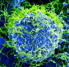
| Colorized scanning electron micrograph of Ebola virus particles (green) found both as extracellular particles and budding particles from a chronically infected African Green Monkey Kidney cell (blue); 20,000x magnification | |
| Virus classification | |
| (unranked): | Virus |
| Realm: | Riboviria |
| Kingdom: | Orthornavirae |
| Phylum: | Negarnaviricota
|
| Class: | Monjiviricetes |
| Order: | Mononegavirales |
| Family: | Filoviridae |
| Genus: | Ebolavirus |
| Species: | Zaire ebolavirus
|
Zaire ebolavirus, more commonly known as Ebola virus (
Ebola virus and its genus were both originally named for Zaire (now the Democratic Republic of the Congo), the country where it was first described,[1] and was at first suspected to be a new "strain" of the closely related Marburg virus.[5][6] The virus was renamed "Ebola virus" in 2010 to avoid confusion. Ebola virus is the single member of the species Zaire ebolavirus, which is assigned to the genus Ebolavirus, family Filoviridae, order Mononegavirales. The members of the species are called Zaire ebolaviruses.[1][7] The natural reservoir of Ebola virus is believed to be bats, particularly fruit bats,[8] and it is primarily transmitted between humans and from animals to humans through body fluids.[9]
The EBOV genome is a single-stranded RNA, approximately 19,000
Because of its high
Structure

.jpg)
EBOV carries a
The overall shape of the virions after purification and visualization (e.g., by
Genome
Each virion contains one molecule of linear, single-stranded, negative-sense RNA, 18,959 to 18,961 nucleotides in length.[20] The 3′ terminus is not polyadenylated and the 5′ end is not capped. This viral genome codes for seven structural proteins and one non-structural protein. The gene order is 3′ – leader – NP – VP35 – VP40 – GP/sGP – VP30 – VP24 – L – trailer – 5′; with the leader and trailer being non-transcribed regions, which carry important signals to control transcription, replication, and packaging of the viral genomes into new virions. Sections of the NP, VP35 and the L genes from filoviruses have been identified as endogenous in the genomes of several groups of small mammals.[21][22][23]

It was found that 472 nucleotides from the 3' end and 731 nucleotides from the 5' end are sufficient for replication of a viral "minigenome", though not sufficient for infection.[18] Virus sequencing from 78 patients with confirmed Ebola virus disease, representing more than 70% of cases diagnosed in Sierra Leone from late May to mid-June 2014,[24][25] provided evidence that the 2014 outbreak was no longer being fed by new contacts with its natural reservoir. Using third-generation sequencing technology, investigators were able to sequence samples as quickly as 48 hours.[26] Like other RNA viruses,[24] Ebola virus mutates rapidly, both within a person during the progression of disease and in the reservoir among the local human population.[25] The observed mutation rate of 2.0 x 10⁻³ substitutions per site per year is as fast as that of seasonal influenza.[27]
| Symbol | Name | UniProt | Function |
|---|---|---|---|
| NP | Nucleoprotein | P18272 | Wraps genome for protection from nucleases and innate immunity. |
| VP35 | Polymerase cofactor VP35 | Q05127 | Polymerase cofactor; suppresses innate immunity by binding RNA. |
| VP40 | Matrix protein VP40 | Q05128 | Matrix. |
| GP | Envelope glycoprotein | Q05320 | Cleaved by host furin into GP1/2 to form envelope with spikes. Also makes shed GP as a decoy. |
| sGP | Pre-small/secreted glycoprotein | P60170 | Shares ORF with GP. Cleaved by host furin into sGP (anti-inflammatory) and delta-peptide (viroporin). |
| ssGP | Super small secreted glycoprotein | Q9YMG2 | Shares ORF with GP; created by mRNA editing. Unknown function. |
| VP30 | Hexameric zinc-finger protein VP30 | Q05323 | Transcriptional activator. |
| VP24 | Membrane-associated protein VP24 | Q05322 | Blocks IFN-alpha/beta and IFN-gamma signaling. |
| L | RNA-directed RNA polymerase L | Q05318 | RNA replicase .
|
Entry

There are two candidates for host cell entry proteins. The first is a cholesterol transporter protein, the host-encoded Niemann–Pick C1 (
When cells from
The second candidate is TIM-1 (a.k.a.
Replication

Being acellular, viruses such as Ebola do not replicate through any type of cell division; rather, they use a combination of host- and virally encoded enzymes, alongside host cell structures, to produce multiple copies of themselves. These then self-assemble into viral
These viral proteins are processed: a glycoprotein precursor (GP0) is cleaved to GP1 and GP2, which are then heavily glycosylated using cellular enzymes and substrates. These two molecules assemble, first into heterodimers, and then into trimers to give the surface peplomers. Secreted glycoprotein (sGP) precursor is cleaved to sGP and delta peptide, both of which are released from the cell. As viral protein levels rise, a switch occurs from translation to replication. Using the negative-sense genomic RNA as a template, a complementary +ssRNA is synthesized; this is then used as a template for the synthesis of new genomic (-)ssRNA, which is rapidly encapsidated. The newly formed nucleocapsids and envelope proteins associate at the host cell's plasma membrane; budding occurs, destroying the cell.[citation needed]
Ecology
Ebola virus is a zoonotic pathogen. Intermediary hosts have been reported to be "various species of fruit bats ... throughout central and sub-Saharan Africa". Evidence of infection in bats has been detected through molecular and serologic means. However, ebolaviruses have not been isolated in bats.[8][38] End hosts are humans and great apes, infected through bat contact or through other end hosts. Pigs in the Philippines have been reported to be infected with Reston virus, so other interim or amplifying hosts may exist.[38] Ebola virus outbreaks tend to occur when temperatures are lower and humidity is higher than usual for Africa.[39] Even after a person recovers from the acute phase of the disease, Ebola virus survives for months in certain organs such as the eyes and testes.[40]
Ebola virus disease
Zaire ebolavirus is one of the four ebolaviruses known to cause disease in humans. It has the highest
, where he recorded the first clinical description of the disease in his daily log:The illness is characterized with a high temperature of about 39°C, hematemesis, diarrhea with blood, retrosternal abdominal pain, prostration with "heavy" articulations, and rapid evolution death after a mean of three days.[42]
Since the first recorded clinical description of the disease during 1976 in Zaire, the recent Ebola outbreak that started in March 2014, in addition, reached epidemic proportions and has killed more than 8000 people as of January 2015. This outbreak was centered in West Africa, an area that had not previously been affected by the disease. The toll was particularly grave in three countries: Guinea, Liberia, and Sierra Leone. A few cases were also reported in countries outside of West Africa, all related to international travelers who were exposed in the most affected regions and later showed symptoms of Ebola fever after reaching their destinations.[43]
The severity of the disease in humans varies widely, from rapid fatality to mild illness or even asymptomatic response.[44] Studies of outbreaks in the late twentieth century failed to find a correlation between the disease severity and the genetic nature of the virus. Hence the variability in the severity of illness was suspected to correlate with genetic differences in the victims. This has been difficult to study in animal models that respond to the virus with hemorrhagic fever in a similar manner as humans, because typical mouse models do not so respond, and the required large numbers of appropriate test subjects are not easily available. In late October 2014, a publication reported a study of the response to a mouse-adapted strain of Zaire ebolavirus presented by a genetically diverse population of mice that was bred to have a range of responses to the virus that includes fatality from hemorrhagic fever.[45]
Vaccine
In December 2016, a study found the
History and nomenclature

Ebola virus was first identified as a possible new "strain" of
In 1998, the virus name was changed to "Zaire Ebola virus"[51][52] and in 2002 to species Zaire ebolavirus.[53][54] However, most scientific articles continued to refer to "Ebola virus" or used the terms "Ebola virus" and "Zaire ebolavirus" in parallel. Consequently, in 2010, a group of researchers recommended that the name "Ebola virus" be adopted for a subclassification within the species Zaire ebolavirus, with the corresponding abbreviation EBOV.[1] Previous abbreviations for the virus were EBOV-Z (for "Ebola virus Zaire") and ZEBOV (for "Zaire Ebola virus" or "Zaire ebolavirus"). In 2011, the ICTV explicitly rejected a proposal (2010.010bV) to recognize this name, as ICTV does not designate names for subtypes, variants, strains, or other subspecies level groupings.[55] At present, ICTV does not officially recognize "Ebola virus" as a taxonomic rank, but rather continues to use and recommend only the species designation Zaire ebolavirus.[56] The prototype Ebola virus, variant Mayinga (EBOV/May), was named for Mayinga N'Seka, a nurse who died during the 1976 Zaire outbreak.[1][57][58]
The name Zaire ebolavirus is derived from Zaire and the taxonomic suffix ebolavirus (which denotes an ebolavirus species and refers to the Ebola River).[1] According to the rules for taxon naming established by the International Committee on Taxonomy of Viruses (ICTV), the name Zaire ebolavirus is always to be capitalized, italicized, and to be preceded by the word "species". The names of its members (Zaire ebolaviruses) are to be capitalized, are not italicized, and used without articles.[1]
Virus inclusion criteria
A virus of the genus Ebolavirus is a member of the species Zaire ebolavirus if:[1]
- it is endemic in the Democratic Republic of the Congo, Gabon, or the Republic of the Congo
- it has a genome with two or three gene overlaps(VP35/VP40, GP/VP30, VP24/L)
- it has a genomic sequence that differs from the type virusEBOV/May by less than 30%
Evolution
Zaire ebolavirus diverged from its ancestors between 1960 and 1976.
A recombination event between Zaire ebolavirus lineages likely took place between 1996 and 2001 in wild apes giving rise to recombinant progeny viruses.[61] These recombinant viruses appear to have been responsible for a series of outbreaks among humans in Central Africa in 2001–2003.[61]
Zaire ebolavirus – Makona variant caused the 2014 West Africa outbreak.[62] The outbreak was characterized by the longest instance of human-to-human transmission of the viral species.[62] Pressures to adapt to the human host were seen at this time, however, no phenotypic changes in the virus (such as increased transmission, increased immune evasion by the virus) were seen.[citation needed]
In literature
- Alex Kava's 2008 crime novel, Exposed, focuses on the virus as a serial killer's weapon of choice.[63]
- William Close's 1995 Ebola: A Documentary Novel of Its First Explosion and 2002 Ebola: Through the Eyes of the People focused on individuals' reactions to the 1976 Ebola outbreak in Zaire.[64][65][66][67]
- The Hot Zone: A Terrifying True Story: A 1994 best-selling book by Richard Preston about Ebola virus and related viruses, including an account of the outbreak of an Ebolavirus in primates housed in a quarantine facility in Reston, Virginia, USA[68]
- Tom Clancy's 1996 novel, Executive Orders, involves a Middle Eastern terrorist attack on the United States using an airborne form of a deadly Ebola virus named "Ebola Mayinga".[69][70]
References
- ^ PMID 21046175.
- PMID 25648530.
- ^ Ebola virus disease (Report). World Health Organization. Retrieved 6 June 2019.
- ^ "Ebola virus disease outbreak". World Health Organization. Retrieved 4 December 2016.
- ^ S2CID 33060636.
- ^ S2CID 3092094.
- ^ WHO. "Ebola virus disease".
- ^ a b Quammen, David (30 December 2014). "Insect-Eating Bat May Be Origin of Ebola Outbreak, New Study Suggests". news.nationalgeographic.com. Washington, DC: National Geographic Society. Archived from the original on 31 December 2014. Retrieved 30 December 2014.
- New York Times. Retrieved 27 October 2014.
- PMID 23383374.
- ^ "Ebola virus disease Fact sheet N°103". World Health Organization. March 2014. Retrieved 12 April 2014.
- ISBN 978-0080575483.
- ISBN 978-0-9545232-3-7.
- )
- PMID 20198110.
- PMID 26024394.
- PMID 27190508.
- ^ ISBN 978-1904933496.[page needed]
- ^ Hillman, H. (1991). The Case for New Paradigms in Cell Biology and in Neurobiology. Edwin Mellen Press.
- ^ Zaire ebolavirus isolate H.sapiens-wt/GIN/2014/Makona-Kissidougou-C15, complete genome, GenBank
- PMID 20569424.
- PMID 20686665.
- PMID 25237605.
- ^ a b Richard Preston (27 October 2014). "The Ebola Wars". The New Yorker. New York: Condé Nast. Retrieved 20 October 2014.
- ^ PMID 25214632.
- PMID 25951262.
- S2CID 20759532.
- ^ PMID 21866103.
- Amanda Schaffer (16 January 2012). "Key Protein May Give Ebola Virus Its Opening". The New York Times.
- ^ PMID 21866101.
- Amanda Schaffer (16 January 2012). "Key Protein May Give Ebola Virus Its Opening". The New York Times.
- S2CID 26888076.
- PMID 22395071.
- PMID 21536871.
- ^ Biomarker Database. Ebola virus. Korea National Institute of Health. Archived from the original on 22 April 2008. Retrieved 31 May 2009.
- PMID 20862315.
- PMID 24093048.
- )
- PMID 25699183.
- ^ S2CID 4657264.
- PMID 25210981.
- ^ "Clinical care for survivors of Ebola virus disease" (PDF). World Health Organization. 2016. Retrieved 4 December 2016.
- ^ Isaacson M, Sureau P, Courteille G, Pattyn, SR. "Clinical Aspects of Ebola Virus Disease at the Ngaliema Hospital, Kinshasa, Zaire, 1976". European Network for Diagnostics of "Imported" Viral Diseases (ENIVD). Archived from the original on 4 August 2014. Retrieved 24 June 2014.
- ^ Bardi, Jason Socrates. "Death Called a River". The Scripps Research Institute. Retrieved 9 October 2014.
- ^ name: S. Reardan.; N Engl. J Med. (2014) " The first nine months of the epidemic and projection, Ebola virus disease in west Africa". archive of Ebola Response Team. 511(75.11):520
- New York Times. Retrieved 30 October 2014.
- PMID 25359852.
- PMID 28017403.
- ^ Berlinger, Joshua (22 December 2016). "Ebola vaccine gives 100% protection, study finds". CNN. Retrieved 27 December 2016.
- ^ "First FDA-approved vaccine for the prevention of Ebola virus disease, marking a critical milestone in public health preparedness and response". U.S. Food and Drug Administration (FDA). 19 December 2019. Archived from the original on 20 December 2019. Retrieved 19 December 2019.
- ^ Brown, Rob (18 July 2014). "The virus detective who discovered Ebola". BBC News.
- S2CID 19368457.
- ISBN 978-0123702005.
- S2CID 13229117.
- ISBN 978-0123702005.
- S2CID 43887711.
- ^ "Replace the species name Lake Victoria marburgvirus with Marburg marburgvirus in the genus Marburgvirus". Archived from the original on 5 March 2016. Retrieved 31 October 2014.
- ^ International Committee on Taxonomy of Viruses. "Virus Taxonomy: 2013 Release".
- PMID 15681442.
- S2CID 34249020.
- ^ PMID 23255795.
- ^ S2CID 9873900.
- ^ PMID 17942693
- ^ a b "Outbreaks Chronology: Ebola Virus Disease". Ebola Hemorrhagic Feve. CDC. 2 August 2017. Retrieved 11 November 2017.
- ISBN 978-0778325574. Retrieved 7 November 2021.
- OCLC 32753758. At Google Books.
- ^ Grove, Ryan (2 June 2006). More about the people than the virus. Retrieved 17 September 2014.
{{cite book}}:|website=ignored (help) - OCLC 49193962. At Google Books.
- ^ Pink, Brenda (24 June 2008). "A fascinating perspective". Review of Close, William T., Ebola: Through the Eyes of the People. Retrieved 17 September 2014.
- OCLC 32052009.
- OCLC 34878804.
- ^ Stone, Oliver (2 September 1996). "Who's That in the Oval Office?". Books News & Reviews. The New York Times Company. Archived from the original on 10 April 2009. Retrieved 10 September 2014.
Further reading
- Pacheco, Daniela Alexandra de Meneses Rocha; Rodrigues, Acácio Agostinho Gonçalves; Silva, Carmen Maria Lisboa da (October 2016). "Ebola virus – from neglected threat to global emergency state". Revista da Associação Médica Brasileira. 62 (5): 458–467. PMID 27656857.
External links
- Ebolavirus molecular biology
- Ebolavirus proteins (PDB-101)
- ICTV Files and Discussions – Discussion forum and file distribution for the International Committee on Taxonomy of Viruses Archived 7 October 2011 at the Wayback Machine
- Genomic data on Ebola virus isolates and other members of the family Filoviridae
- ViralZone: Ebola-like viruses – Virological repository from the Swiss Institute of Bioinformatics
- Virus Pathogen Resource: Ebola Portal – Genomic and other research data about Ebola and other human pathogenic viruses
- The Ebola Virus 3D model of the Ebola virus, prepared by Visual Science, Moscow.
- FILOVIR – scientific resources for research on filoviruses Archived 30 July 2020 at the Wayback Machine
- "'Zaire ebolavirus'". NCBI Taxonomy Browser. 186538.
- "'Ebola virus sp.'". NCBI Taxonomy Browser. 205488.