Chandelier cell
| Chandelier cell | |
|---|---|
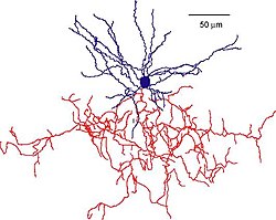
 Reconstruction of a mouse chandelier cell. Soma and dendrites are labeled in blue, axon arbor in red. (Woodruff & Yuste, 2008, PLoS Biology)[1] | |
| Anatomical terms of neuroanatomy |

An action potential in a pyramidal neuron (cell 1) elicits a spike in a chandelier cell (2) via a strong connection, in turn evoking a third-order spike in a downstream pyramidal cell (3). This spike results in a trisynaptic EPSP being recorded in a postsynaptic pyramidal cell (cell 4, event A). At the same time, cell 3 drives both a basket cell (5) and chandelier cell (6) to threshold. The basket cell evokes a hyperpolarizing IPSP on the postsynaptically recorded pyramidal cell (cell 4, event B), four synapses removed from the original spike. The spiking chandelier cell (6) triggers yet another pyramidal neuron to fire (7), which produces an EPSP on the recorded neuron (cell 4, event C), five synapses away from the original spike. The result seen in the postsynaptic pyramidal neuron (cell 4) is a delayed EPSP-IPSP-EPSP sequence (events A, B, and C), traveling through three, four, and five synapses respectively. Molnár et al. propose[2] that polysynaptic pathways similar to this one can be activated by a single action potential in a cortical pyramidal cell.
Chandelier cells or chandelier neurons are a subset of
axonal initial segment of pyramidal neurons, near the site where action potential is generated.[6] It is believed that they provide inhibitory input to the pyramidal neurons, but there is data showing that in some circumstances the GABA from chandelier neurons could be excitatory. [7]
The axon cartridges formed by chandelier cells are one of the synapse types that show the most dramatic changes during normal adolescence,[8] and could potentially be relevant to the adult onset of psychiatric disease. Furthering this link, in schizophrenia, scientists have observed changes in their form and functionality, such as 40% decrease in the axon terminal density.[9]
See also
List of distinct cell types in the adult human body
References
External links
Wikimedia Commons has media related to Chandelier cell.
- SRF interviews David Lewis - an interview touching on the GABAergic neuronal dysfunction in schizophrenia and the role of the chandelier cells.
- NIF Search - Chandelier Cell via the Neuroscience Information Framework
- Cortical Development - images of chandelier neurons and information on their developmental changes. Translational Neuroscience Program at the University of Pittsburgh.
- How chandelier cells light up human thought - A type of brain cell called a chandelier neuron might be what gives us the edge over other mammals in thought and language, New Scientist, 3 September 2008
