
Johan Sebastiaan Ploem
Johan Sebastiaan Ploem | |
|---|---|
| Born | 25 August 1927 University of Leiden |
| Thesis | Enkele methoden voor toxiciteitsonderzoek met behulp van weefselkweekcellen (1967) |
| Doctoral advisor | Jos van der Meer |
| Website | ploem-reflection-contrast-microscopy |
Johan Sebastiaan Ploem
Early life and education
Ploem was born on 25 August 1927 in Sawahlunto in West Sumatra,[4] then part of the Dutch East Indies. At 2 years old his family moved to the Netherlands, where he remained the rest of his youth.[2]
Ploem received an MD at
Scientific career
Ploem has been employed by a number of academic institutions, including the
Fluorescence microscopy
Ploem is most well known for inventing the epi-illumination cube used in fluorescence microscopy.[6][7]
Around 1962 Ploem started work in collaboration with Schott on the development of dichroic beam splitters for reflection of blue and green light for fluorescence microscopy using epi illumination. At the time of his first publication[8] on fluorescence microscopy using epi illumination with narrow-band blue and green light, he was not aware of the development of a dichroic beam splitter for UV excitation with incident light by Brumberg and Krylova.
Ploem's prototype fluorescence epi-illuminators and microscopes form a part of the permanent exposition of the Dutch National Museum for the history of Science and Medicine.[9]
Reflection contrast microscopy
In 1973, at the Second Conference on Mononuclear Phagocytes held in Leiden, Ploem introduced an improvement to Interference Reflection Microscopy (IRM), which he called Reflection Contrast Microscopy (RCM). He also wrote a book chapter in the associated conference proceeding edited by Ralph van Furth.[10] The improvement is the addition of crossed polarizers and a so-called "anti-flex objective", the combination of which further reduces stray light in an IRM microscope, allowing even better interference contrast.[11] RCM is more commonly known as Reflection Contrast Interference Microscopy (RICM) today.[12][13]
Honors and awards
Various awards and honors have been bestowed on Ploem:[3]
- Fellow of the Papanicolaou Cancer Research Institute, Miami, Florida, 1977
- Fellowship to the Institute for Cell Analysis at the University of Miami, Florida, 1979
- C. E. Alken Foundation award, co-recipient, Switzerland, 1982
- Ernst Abbe Medal and Award of the New York Microscopical Society, 1998[3]
- Erica Wachtel medal from the British Society for Clinical Cytology, 1993 (held the Erica Wachtel Medal Lecture)
- The first Honorary member of the International Society for Analytical Cytology, 1993
- Honorary fellow of the Royal Microscopical Society (FRMS), 1976, Oxford[14]
- Honorary fellow of the New York Microscopical Society
- Honorary fellow of the Polish Society of Surgery
- Honorary fellow of the German Society of Surgery
- In 1994, the European Society for Analytical Cellular Pathology established a Conference Keynote "Ploem" Lecture for invited scientists at its future general meetings
- The International Society of Analytical Cytology invited Professor Ploem to present its inaugural "Robert Hooke" lecture.
- In 1995, he was invited by the Royal Microscopical Society to give the inaugural CYTO lecture.
Digital painting
Ploem started painting as a small boy and was educated in drawing and painting in
In the last years of his activities at the faculty of medicine at Leiden University, he concentrated on research in image analysis. He was asked to participate in a European project with the aim of automating cancer cell recognition using computer analysis. It concerned a collaborative project with the German optical company Leitz/Leica Microsystems, and the Institute for Mathematical Morphology in Fontainebleau, France. Together with a team, Professor Jean Serra at this institute had developed an image analysis method, now internationally known as mathematical morphology. With his experience as an analogue painter, Ploem saw the possibility of also applying the methods of mathematical morphology to the creation of digital art.
At the International Symposium on Mathematical Morphology in Amsterdam (1998), Ploem presented a paper on the creation of computer graphics with Mathematical Morphology, using for the first time, the transforming algorithms from the Fontainebleau group for the creation of digital art. He wrote about it in the chapter of a book (
His first digital graphics of nature scenes were shown in his exposition at a regional art centre in the Pyrenees (Ossega, June 1997).[citation needed]
He was invited for a symposium on Art et Science at the
-
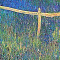 Meadow near Sauto, Pyrenees, France
 -
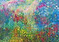 In the valley of the Eyne, Pyrenees
 -
 David and Goliath (multiple image transformations of a romanesque painting in the Sant Climent church in Taull)


References
- ^ "Ploem-reflection-contrast-microscopy.com". Archived from the original on 2023-02-16.
- ^ a b c "Johan Sebastiaan Ploem (1927–Present)". micro.magnet.fsu.edu. Retrieved 2023-11-11.
- ^ a b c d e "Bas Ploem (Cytometry Volume 10)". cyto.purdue.edu. Purdue University Cytometry Laboratories. 1998. Retrieved 2023-11-11.
- ^
"Johan Sebastiaan Ploem (Bas)". hoogleraren.universiteitleiden.nl. Universiteit Leiden. Retrieved 2023-11-10.
Geboren: 1927-08-25 te Sawahlunto, Nederlands-Indië
- ^
Ploem, Johan Sebastiaan (1967). Enkele methoden voor toxiciteitsonderzoek met behulp van weefselkweekcellen (Ph.D.). OCLC 898812117.
- ^ "Pioneers in the Optical Sciences". micro.magnet.fsu.edu. Retrieved 2023-11-11.
- ^ www
.olympus-lifescience .com /en /microscope-resource /primer /techniques /fluorescence /reflectlightpaths - ^ Ploem, J. S. (1967). "Die Möglichkeit der Auflichtfluoreszenzmethoden bei Untersuchungen von Zellen in Durchströmungskammern und Leightonröhren, Xth Symposium d. Gesellschaft f. Histochemie". Acta Histochemica. Supplementband. 7: 339–343. .
- ^ http://journal.sciencemuseum.ac.uk/browse/issue-08/museum-theme-the-quest-for/rijksmuseum-boerhaave
- OCLC 2701405.
- S2CID 73431526.
- PMID 19947618.
- ^
Limozin, Laurent; Sengupta, Kheya (2009-11-03). "Quantitative Reflection Interference Contrast Microscopy (RICM) in Soft Matter and Cell Adhesion". ChemPhysChem. 10 (16). Wiley: 2752–2768. PMID 19816893.
- ^
"Current RMS Honorary Fellows". rms.org.uk. Royal Microscopical Society. Retrieved 2023-11-10.
1976 Professor J S Ploem Hon FRMS
- ^
Heijmans, Henk J.A.M.; Roerdink, Jos (1998-05-31). Mathematical Morphology and its Applications to Image and Signal Processing. Dordrecht: Kluwer (Springer Science & Business Media). p. 355. OCLC 39348037.
External links
- Official website

- Website Leiden Professors (in Dutch)
| International | |
|---|---|
| National | |
| Academics | |
| People | |
| Other | |