Peripherin
| PRPH | |||
|---|---|---|---|
| Identifiers | |||
Gene ontology | |||
| Molecular function | |||
| Cellular component | |||
| Biological process | |||
| Sources:Amigo / QuickGO | |||
Ensembl | |||||||||
|---|---|---|---|---|---|---|---|---|---|
| UniProt | |||||||||
| RefSeq (mRNA) | |||||||||
| RefSeq (protein) |
| ||||||||
| Location (UCSC) | Chr 12: 49.29 – 49.3 Mb | Chr 15: 98.95 – 98.96 Mb | |||||||
| PubMed search | [3] | [4] | |||||||
| View/Edit Human | View/Edit Mouse |
Peripherin is a type III
History
Peripherin, first named such in 1984, was also known as 57 kDa neuronal intermediate filament prior to 1990. In 1987, a second distinct peripherally located retinal rod protein was also given the name peripherin. To distinguish between the two, this second protein is referred to peripherin 2 or peripherin/RDS (retinal degeneration slow) for its location and role in retinal disease.[8]
Structure and properties
Peripherin was discovered as being the major intermediate filament in
Peripherin, unlike keratin IFs, can self-assemble and exist as homopolymers (see
In addition to the main species of peripherin, 57 kDa, two other forms have been identified in mice: Per 61 and Per 56. These two alternatives are both made by alternative splicing. Per 61 is created by introducing a 32 amino acid insertion within coil 2b of the α-helical rod domain of peripherin. Per 56 is made by a receptor on exon 9 of the peripherin gene transcript which induces a frameshift and replacement of a 21 amino acid sequence in the C-terminal found on the dominant 57 form with a new 8 amino acid sequence. The functions of these two alternative forms of peripherin are unknown. Per 57 and 56 are normally co-expressed, whereas Per 61 is not found in normal peripherin expression in adult motor neurons.[12]
Tissue distribution
Peripherin is widely expressed in the cell body and axons of neurons in the
A comparison of peripherin expression in the posterior and lateral hypothalamus in mice showed a sixty-fold higher expression in the posterior hypothalamus. This higher expression is due to the presence of peripherin in the tuberomammillary neurons of the mouse posterior hypothalamus.[13]
Function
The diverse properties of intermediate filaments, compared with the conserved microtubule and actin filament proteins, could be responsible for the distinguishing molecular shapes of different cell types. In nerve cells, for example, the expressions of different types of IFs relates to the change in shape during development. Early stages of development in neurons is marked by the outgrowth of
The exact function of peripherin is unknown. Expression of peripherin in development is greatest during the axonal growth phase and decreases postnatally, which suggests a role in neurite elongation and axonal guidance during development. Expression is also increased after axonal injury, such as peripheral

Gene (PRPH)
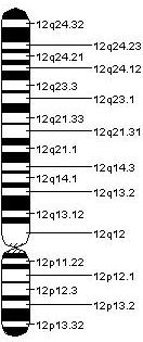
The complete sequence of the human (GenBank L14565), rat (GenBank M26232) and mouse (EMBL X59840) peripherin genes (PRPH) have been reported and complementary DNAs (cDNA) thus far described are those for rat, mouse and Xenopus peripherin.[8] The use of a mouse cDNA probe during the in situ hybridization procedure allowed for the localization of the PRPH gene to the E-F region of mouse chromosome 15 and the q12-q13 region of human chromosome 12.[6]
The overall structure of the peripherin gene is nine
Regulatory mechanisms
The proinflammatory cytokines,
Potential role in the pathogenesis of amyotrophic lateral sclerosis
Protein and neurofilamentous aggregates are characteristic of patients with amyotrophic lateral sclerosis, a progressive, fatal neurodegenerative disease. Spheroids, specifically, which are protein aggregates of neuronal intermediate filaments, have been found in patients with amyotrophic lateral sclerosis. Peripherin has been found in such spheroids in conjunction with other neurofilaments in other neuronal diseases, thus suggesting that peripherin may play a role in the pathogenesis of amyotrophic lateral sclerosis.[7]
Alternative splicing
An alternatively spliced mouse peripherin variant was identified that includes intron 4, a region that is spliced out of the abundant peripherin forms. Because of the change in reading frame, this variant produces a larger form of peripherin (Per61). In human peripherin, the inclusion of introns 3 and 4, regions that are similarly spliced out of the abundant peripherin protein forms, results in the generation of a truncated peripherin protein (Per28). In both cases, an antibody specific to a peptide coded by the intron regions stained the filamentous inclusions of in tissues affected by amyotrophic lateral sclerosis. These studies suggest that such alternative splicing could play a role in the disease and lend themselves to further investigation.[7]
Mutations
Experiments examining peripherin overexpression in mice have suggested that PRPH mutations play a role in the pathogenesis of amyotrophic lateral sclerosis, with more recent studies investigating the prevalence of such mutations in humans. Though many
The second variant consisted of an amino acid substitution from
Other clinical significance
Peripherin may be involved in the pathology of insulin-dependent diabetes mellitus (or
Peripherin can also play a role in the definitive diagnosis of
Potential applications
Possible involvement of intermediate filaments such as peripherin in neurodegenerative diseases is currently being investigated. Interactions between intermediate filaments and other proteins are also being pursued. Peripherin has been shown to associate with protein kinase Cε, inducing its aggregation and leading to increased
References
- ^ a b c GRCh38: Ensembl release 89: ENSG00000135406 – Ensembl, May 2017
- ^ a b c GRCm38: Ensembl release 89: ENSMUSG00000023484 – Ensembl, May 2017
- ^ "Human PubMed Reference:". National Center for Biotechnology Information, U.S. National Library of Medicine.
- ^ "Mouse PubMed Reference:". National Center for Biotechnology Information, U.S. National Library of Medicine.
- ^ "Entrez Gene: Peripherin".
- ^ PMID 1378416.
- ^ PMID 19587456.
- ^ a b c d Vale, Ronald; Kreis, Thomas (1999). Guidebook to the Cytoskeletal and Motor Proteins (2nd ed.). Sambrook & Tooze Partnership.
- ^ S2CID 15737750.
- ^ PMID 7979242.
- ^ PMID 16505169.
- ^ PMID 17045786.
- ^ PMID 18294400.
- S2CID 31835055.
- PMID 7806235.
- PMID 15322088.
- S2CID 43439366.
- PMID 20495540.
- PMID 18408015.
External links
- Peripherin at the U.S. National Library of Medicine Medical Subject Headings (MeSH)
