
Ticks of domestic animals

Ticks of domestic animals directly cause poor health and loss of production to their hosts. Ticks also transmit numerous kinds of viruses, bacteria, and protozoa between domestic animals.[1] These microbes cause diseases which can be severely debilitating or fatal to domestic animals, and may also affect humans. Ticks are especially important to domestic animals in tropical and subtropical countries, where the warm climate enables many species to flourish. Also, the large populations of wild animals in warm countries provide a reservoir of ticks and infective microbes that spread to domestic animals. Farmers of livestock animals use many methods to control ticks, and related treatments are used to reduce infestation of companion animals.
Variety of ticks affecting domestic animals

Ticks are invertebrate animals in the phylum
Typical ticks of domestic animals
Amblyomma and Rhipicephalus ixodid ticks


Amblyomma species are widespread on domestic animals throughout tropical and subtropical regions. Typical Amblyomma species are: Amblyomma americanum, the lone star tick of the Southern and Eastern USA; Am. cajennense, the Cayenne tick of South America and Southern USA; Amblyomma variegatum, the bont tick of Africa and the Caribbean (see Gallery below for photograph of female and male). A typical Rhipicephalus species is Rhipicephalus sanguineus, the tropical dog tick, specialized to feed only on dogs. It is distributed globally throughout the warm countries, wherever humans with their dogs live. Typical Rhipicephalus species that feed on cattle in Africa are R. appendiculatus, the brown ear-tick, and R. evertsi, the red-legged tick. Rhipicephalus (Boophilus) microplus (or now simply Rhipicephalus microplus) is the most important tick of cattle in many tropical and subtropical countries to which it spread from Southeast Asia on transported cattle.
Boophilid ticks, a subgenus within Rhipicephalus

These ticks, commonly known as cattle ticks or blue ticks, have a highly characteristic morphology and one-host lifecycle. They have high specificity for cattle as hosts and their morphological characteristics used for identification are less distinct than those of three-host rhipicephalids such as R. appendiculatus. They are economically important to the cattle-rearing industry by causing direct parasitic losses and by transmission of microbes. In addition to Rhipicephalus microplus, species of most importance to domestic animals are R. annulatus, which is widespread in tropical and subtropical countries, and R. decoloratus which occurs in Africa.[citation needed]
One-host lifecycle of R. microplus

These ticks are adapted to the advantages of specialising to feed on cattle and with all the feeding stages occurring on one individual host in a rapid sequence. They also can survive by feeding on deer or some wild bovid hosts. Infestation starts when larvae on vegetation attach to a new host. When a larva feeds, it molts at the site where it feeds and emerges as a nymph. The nymph feeds at the same site or close by and molts where it feeds. It emerges from molt as either an adult female or male. The female's single large blood meal is converted into a batch of 2000 eggs. The males take several small meals of blood to support their repeated attempts at mating. The molts are rapid and the next stage remains in the hair coat to start feeding again. The combined feeding and molting periods take about 21 days. The engorged female drops from the host, hides under leaf litter on soil surface, lays one batch of eggs, and then dies. When eggs hatch, the larvae crawl up grass stems and wait until they can attach to passing cattle.[citation needed]
Hyalomma ticks

This genus contains many species of hard ticks important to domestic animals in hot, dry regions in Africa, the Mediterranean basin, the Middle East, Pakistan, India,[7] and through to China. Typical species are Hyalomma anatolicum, Hy. rufipes, Hy. truncatum, and Hy. detritum, which feed as adults on cattle, sheep, and goats. Hyalomma dromedarii is specialized to feed on dromedary camels. Hyalomma ticks are adapted to live in regions with large seasonal variation of temperature and low rainfall. Diapause is an important mechanism to adjust to these climates. Another adaptation is to have a lifecycle within one species that can be two-host or three-host. For example: Hy. anatolicum may feed on a hare, molt on the hare, and feed again on the same individual hare, detach and molt to an adult and then feed on a cow - that is a two-host lifecycle. Or it may feed as a larva on a gerbil, then as a nymph on a cow, and then as an adult on another cow in a three-host lifecycle. Furthermore, this tick commonly feeds as a three-host tick with larvae, nymphs, and adults feeding on separate individual dairy cows confined to cattle housing in zero-grazing systems.[citation needed]
Argas and Ornithodoros soft ticks

Argas persicus, the fowl tick, is a major pest of poultry birds. The tampan ticks within the Ornithodoros moubata complex of species infest domestic pigs and also feed on humans. Ornithodoros savignyi is often found in large numbers at enclosures where camels and cattle are herded. Many species of argasid soft ticks are adapted to live in the nest or regular resting sites of their hosts, often waiting for months or even years for the host to return and enable the tick to feed. This nest-dwelling behavior is described as endophilic or nidicolous.[citation needed]
Multihost lifecycle of O. moubata

Argasidae soft ticks have different lifecycles from Ixodidae hard ticks, and these are very variable between species.[1] Typically, in Ornithodoros, a larva hatches from an egg laid in the nest or resting place of the host. The larva does not feed, but directly molts into the first nymph stage. This stage feeds, then molts into the next nymph stage. Feeding by soft ticks is generally completed within minutes rather than days, as with hard ticks. Depending on circumstances, four or five nymph stages occur, each progressively larger. Finally, a molt produces an adult female or male. The female takes repeated blood meals that are small compared to a female hard tick. Each blood meal is converted to a small batch of eggs. The male feeds sufficiently to support its mating. The lifecycle of Argas persicus is similar, but the larva feeds on blood of its bird host, remaining attached around 7 days.[citation needed]
Other groups of ticks
Other genera with species that are often of high local importance to domestic animals include the following examples, some of which are illustrated in the gallery below. Ixodes (Ixodes ricinus, the deer tick of Europe; Ixodes scapularis, the black-legged tick of North America; Ixodes holocyclus, the paralysis tick of Australia). Haemaphysalis (Ha. leachii, the yellow dog tick of the tropics). Dermacentor (Dermacentor andersoni, the Rocky Mountain wood tick; Dermacentor variabilis, the American dog tick; D. reticulatus, the ornate dog tick of Europe). D. nitens, the tropical horse tick of the Americas, has a one-host lifecycle similar to the boophilids. Margaropus winthemi, the beady-legged tick, infests horses and cattle in South Africa. The soft tick Otobius megnini, the spinose ear tick, has its nymphs feeding within the ear canal of many species of domestic animals. Adults of Ot. megnini do not feed. This tick occurs in the Americas and has spread to Africa and Asia.[citation needed]
Negative impacts to health
Biting stress and lost production

When a hard tick pierces the skin of its host, initially little or no pain is caused. Later, during the prolonged feeding of ticks, inflammation is caused at the wound, followed by acquired immune reactions in the skin (dermal hypersensitivities types 1 and 4) to the foreign proteins in tick saliva. This defense by the host is generally effective, but at the cost of pruritus (itch) and pain at the feeding site. Infestations of ticks on certain individual animals of a herd of livestock animals can build up to very high levels. This occurs on a minor proportion of individuals in the herd, whilst most individual animals have low infestations. On a herd basis, the accumulated effect of this biting stress can cause
Physical damage

At each feeding site of hard ticks, granuloma and wound healing produce a scar that remains for years after the tick has detached. When the skin of livestock animals is made into leather, these scars remain as blemishes that reduce the value of the leather. Larger ticks cause obstructive and painful damage, such as Amblyomma variegatum adults, which often feed on udders of cattle and reduce suckling by the calves. Hyalomma truncatum adults feed on the feet of sheep and goats, causing lameness. Wounds caused by dense clusters of adult ticks can make the host susceptible to infestation with larvae of flesh-eating myiasis flies, such as the screw-worm, Cochliomyia hominivorax.[10][11]
Poisoning

When ticks feed, they secrete saliva containing powerful enzymes and substances with strong pharmacological properties to maintain flow of blood and reduce host immunity. Sometimes, this causes a poisoning of the host. This is not because of a functional toxin in the sense that snake poison is functional for the snake. However, the result can be various forms of toxaemia caused by a variety of ticks. A moist eczema, sometimes with hair loss (alopecia) known as sweating sickness in cattle is caused by Hyalomma truncatum. Tick paralysis can be life-threatening and is caused in sheep by feeding of Ixodes rubicundus of South Africa. In cattle, paralysis is caused by both Dermacentor andersoni in North America and the Australian paralysis tick, Ixodes holocyclus. I. holocyclus also causes paralysis in dogs and humans.[12][13][14]
Ticks as vectors of disease
Because ticks feed repeatedly and only on blood, and have long lives, they are suitable hosts for many types of microbes, which exploit the ticks for transmission between one domestic animal and another. Ticks are thus known as vectors (transmitters) of microbes. Most of these parasitic relationships are highly developed with a strict biological relationship between the microbe and the tick's gut and salivary glands. However, some microbes, such as Anaplasma marginale and A. centrale, can also be transmitted by biting flies, or by blood on injection needles (iatrogenic transmission). A characteristic of diseases caused by tick-transmitted microbes is that herds or flocks of livestock often acquire effective levels of immune resistance to both the vector ticks and the microbes, so outbreaks of acute disease tend to be rare. This stability is often due to immunity to the microbes developing as a result of survival through early infection from ticks carrying small infective doses of the microbe, the epidemiology of infections with Babesia species of protozoa is a well described example.[15] The ticks are often constantly present and long-lived. Acquisition of immunity may be aided by the protection of antibodies in the mother's colostrum (first milk).
At least one microbe causing disease associated with ticks is not transmitted by the ticks. The skin disease dermatophilosis of cattle, sheep, and goats is caused by the bacterium Dermatophilus congolensis, which is transmitted by simple contagion. When Amblyomma variegatum adult ticks are also feeding and causing a systemic suppression of immunity in the host, then dermatophilosis becomes severe or even fatal.[16]
Viral diseases
The virus of Nairobi sheep disease in East Africa is transmitted by Rhipicephalus ticks. African swine fever is naturally transmitted between wild species of the pig family by feeding of Ornithodoros moubata group ticks. This pattern of transmission can expand to include domestic pigs. However, within groups of domestic pigs, the virus can also be transmitted by contagion. Crimean-Congo hemorrhagic fever virus is transmitted between many mammal species by Hyalomma truncatum, Hyalomma rufipes, and Hyalomma turanicum over a wide area of Africa, Europe, and Asia. In cattle and sheep, it causes mild fever and its main importance is when it spreads to humans (zoonosis) by feeding of the larvae or nymphs of these ticks.[17] There are other viruses transmitted by ticks between wild animals and that have zoonotic importance when humans also become infected. The epidemiological pathways of these viruses can also involve domestic animals, if only by being hosts that add to the size of the tick population. Examples include the viruses that cause Tick-borne encephalitis, and Kyasanur Forest disease.[18]
Bacterial diseases

Protozoal diseases

Babesia bovis protozoa are transmitted by R. microplus and cause
Disease control methods
Treatment with chemical and botanical pesticides against ticks
Synthetic chemical acaricides

Ticks infesting sheep and cattle have been controlled with a wide variety of chemicals ranging from coal tar extracts, arsenic salts, and specific pesticidal chemicals such as DDT for many decades. These are now replaced by various synthetic chemicals of high specificity for acarines and ticks, and farmers frequently rely on treating their animals with these materials as their default method.
Botanical acaricides
Farmers lacking access to, or sufficient cash for, manufactured synthetic acaricides often use various herbal treatments, locally available. Nicotine from treated tobacco leaf is an example, but such unregistered preparations require careful use to avoid poisoning or skin damage. Commercially formulated botanical acaricide may often be available in tropical regions, containing the active ingredient azadirachtin. This is extracted as neem oil from fruits and seeds from the neem tree, Azadirachta indica.[30]
Eradication of ticks
Eradication of ticks, as total removal of all populations of a species over a wide geographical area defined by natural boundaries, has been attempted several times.[31] In the southern states of the USA the tick then known as Boophilus annulatus (Rhipicephalus annulatus) was eradicated for the purpose of control of babesiosis in cattle. The eradication was successful after more than 50 years of control with much emphasis on dipping in chemical acaricides. The tick was eradicated up to the border of USA with Mexico, and a control and quarantine zone remains in place there.[32] Similar efforts were made to eradicate the tick then known as Boophilus microplus (also a Rhipicephalus, Rhipicephalus microplus) from New South Wales, Australia. However, this failed, partly due to the difficulty of maintaining a barrier against invasions from the more favourable areas for the tick in sub-tropical Queensland.[33] Amblyomma variegatum was subject to a multi-country eradication program in the Caribbean area, but it failed for complex economic and political reasons.[34]
Biological and ecological based methods in control of ticks and transmitted microbes
Cattle breeding for disease resistance

Breeding for resistant cattle has been successful for their ability to acquire strong immune resistance to Rhipicephalus microplus following natural exposure to these ticks.[35] Commercial breeds of cattle (examples: Australian Friesian Sahiwal and Australian Milking Zebu) are successful in the relevant environment. Only a few commercial breeds of tick-resistant cattle are available. These breeds were developed under laboratory conditions where bulls were selected for good ability to acquire immune resistance to ticks, and cows were selected for heat tolerance and milk yield. The scope for selecting cattle under farm conditions includes culling those animals persistently with heavy infestations of ticks. This is due to a characteristic of many parasitic infestations where in a population of hosts, a few individual hosts carry heavy infestations, whilst the majority are lightly infested. This is called an overdispersed, or aggregated, distribution; it may be caused by individual variation in immune competence, which has a genetic component.[36]
Pasture management

For farms infested with R. microplus in Australia and South America, rotation of pasture can kill the questing larvae.[37] Pasture management is feasible for control of these one-host ticks because the only stage that quests for hosts on vegetation is the larval stage. Because of their small size, larvae are highly susceptible to dehydration and starvation, so a period without access to hosts of about 2 months can be effective. The nymphs and adults of three-host ticks, such as Amblyomma and Rhipicephalus species, can live for many months to a year or more, respectively, whilst questing on the vegetation. Thus, controlling them by pasture management is usually not practical. In addition, farmers find the priority to provide their stock with good feed often conflicts with the regimens for pasture rotation for tick control.[38] Ticks affecting dogs and other companion animals around private houses are reduced by clearing of vegetation and leaf litter, mowing grass short, and fencing out deer and other wild animals that bring in ticks.
Epidemiological factors
Compared to other arthropods of veterinary and medical importance, most species of ticks are long-lived over their whole lifecycles. Ixodes species in cool, temperate climates typically take one year to develop through each of the three feeding stages. Amblyomma species may also have a three-year lifecycle, and the adults can live off-host, without feeding, for up to 2 years. Ornithodoros and Argas ticks are particularly adapted to wait for their hosts to arrive by being able to survive for years between blood meals as adults, 18 years have been recorded for O. lahorensis.[2][39] Food reserves for survival of off-host ticks include large membrane-bound vesicles of lipid in the digestive cells of their gut. Further adaptations include a thick integument with waxy waterproofing combined with ability to secrete hygroscopic salts from specialized parts of their salivary glands (type 1 acini) out to the exterior of their mouthparts and then suck back in the watery solution that develops around the salty material.[40]

At least some species of ticks have fairly stable populations, with variations in population densities from year to year varying by roughly 10- to 100-fold.[41] The abiotic (environmental) factor that appears to have most influence on tick distribution and abundance is dehydration stress from the combination of high temperatures and low moisture (measured as relative humidity or low saturation deficit, usually resulting from a climate of low rainfall). Computer models to test hypotheses and to predict the distribution of ticks reflect these stresses, using meteorological information where available, or indices of vegetation type as an analogue of temperature and dehydration stress.[42][43] Biotic (host related) factors that influence tick distribution and abundance include the obvious need for the hosts to which the ticks are adapted to be present, and also the defenses that the hosts have against the ticks. During the lifecycle of a three-host tick feeding on its natural host that has acquired immunological resistance to the feeding of the ticks, tick mortality can be high.[44] This mortality is highest for the larvae which are easily killed by the immune reactions in the host's skin. It is lowest in the feeding adults. However, even if the ticks do feed, detach, and moult, the size of newly moulted adults is smaller, and the reproductive capacity of the females is reduced.[45] These biotic factors are likely to produce a pattern of mortality of the ticks that is density-dependent.[43] A high density of ticks attempting to feed induces strong immune resistance in their hosts, but a low density of ticks does not induce such strong resistance.
Ticks combine long life of the stages of that carry pathogenic microbes and long survival of these microbes in specialized niches within the tick, such as within cells of the salivary glands or the gut. In a population of Rhipicephalus or Hyalomma ticks feeding on cattle in which Theileria species of protozoa circulate and cause theileriosis, the ticks act as long-term reservoirs of the protozoans. In addition, some species of protozoans (within the Theileria and Babesia genera), are able to infect ticks even when they exist in the blood of their hosts at such a low level that no signs of disease can be detected. This is known as a carrier state of infection.[46][47] These pathogenic protozoa can be detected circulating in populations of the cattle hosts and tick vectors with only low levels of detectable disease in the cattle caused by the protozoa. Any situation like this - with prevalent infection or infestation but little disease - is called endemic stability.[48] It is possible that this can be exploited for better control of tick related diseases by use of breeds of cattle with good ability to acquire resistance to both the ticks and the protozoans. However, there are commonly situations where the potential benefits of endemic stability to disease are difficult for farmers to use effectively. The farmers may prefer to rely more on direct tick control, and drugs and vaccines against the protozoans.[49][50]
Drug treatment against microbes
Antibiotics with efficacy against bacterial pathogens transmitted by ticks include
Vaccination against microbes and ticks
Vaccination against An. marginale is done using live strains of the cross-reactive An. centrale.[53] Vaccines are available on a commercial basis to immunize cattle against Babesia bovis.[54] This is made by serial infection of calves to attenuate the virulence of the strain of Babesia, followed by splenectomy to produce many of the piroplasm stage in blood, which is then bottled for use. The vaccine is delivered containing the live protozoa to induce immunity without acute disease.[55] Theileria annulata can be grown and attenuated in virulence by means of infecting cell cultures with the schizont stage of the protozoan. This is delivered as a frozen vaccine from which live parasites are thawed out before injection.[56][57] Cattle can be protected against East Coast fever by an infection-and-treatment procedure. Rhipicephalus appendiculatus ticks are infected with Theileria parva under laboratory conditions; theilerial sporozoites are extracted from the ticks and stored in liquid nitrogen; infective doses of the live vaccine are delivered to identified cattle and a few days later a protective dose of antibiotic is delivered to stop the infection from developing into clinical East Coast fever.[58] Vaccines are often highly effective, but the live parasite vaccines have problems of potential contamination with other microbes and induction of a carrier state which may be unwanted. Intensive attempts are made to develop vaccines to control these diseases using a recombinant DNA techniques to synthesize the relevant antigens, but as with vaccines against human malaria this is a difficult technological challenge.[59][60]
A commercial vaccine was developed in Australia against Rh. microplus.[61][62] It acts against a glycoprotein molecule that is exposed on the outer membrane of digestive cells of the gut of feeding ticks. This molecule is synthesized using recombinant DNA technique to make the antigen of the vaccine. Vaccinated cattle develop antibodies circulating in their blood. When the Rh. microplus female ticks engorge with blood, the antibody reacts with the natural antigen in their guts so strongly that digestion is disrupted and the reproductive rate of the ticks is reduced. This vaccine is manufactured in Australia and a closely similar vaccine is manufactured in Cuba.[63]
References
- ^ .
- ^ ISBN 0-19-505910-7
- .
- S2CID 4120611.
- S2CID 38865837.
- ISBN 978-1-4051-1964-1.
- OCLC 19554336.[page needed]
- PMID 16472920.
- S2CID 24594154.
- S2CID 12470638.
- ^ ISBN 978-0-85312-790-1.[page needed]
- PMID 232161.
- S2CID 23861588.
- PMID 24943801.
- S2CID 8624479.
- PMID 8161379.
- ISBN 978-0-19-570506-5.[page needed]
- S2CID 205949828.
- PMID 41420.
- hdl:1874/24456.
- ISBN 978-0-12-521740-8.[page needed]
- PMID 19837514.
- S2CID 11953803.
- PMID 9500178.
- ISBN 978-0-511-55180-2.
- PMID 16497440.
- PMID 2056504.
- S2CID 7624515.
- ISBN 978-0-19-507313-3.
- ^ Handule, Ismail Mohamed; Ketavan, Chitapa; Gebre, Solomon (31 March 2002). "Toxic Effect of Ethiopian Neem Oil on Larvae of Cattle Tick, Rhipicephalus pulchellus Gerstaeker". Agriculture and Natural Resources. 36 (1): 18–22.
- S2CID 206246539.
- ISBN 978-0-8203-2749-5.[page needed]
- PMID 9024884.
- ISBN 978-1-4269-4453-6.[page needed]
- PMID 5068812.
- S2CID 10950302.
- PMID 5103591.
- PMID 7436925.
- PMID 3904345.
- ISBN 978-0-511-55180-2.
- PMID 23327204.
- S2CID 10131322.
- ^ ISBN 978-0-511-55180-2.
- .
- S2CID 86525933.
- S2CID 20471003.
- ProQuest 750568336.
- PMID 11423357.
- PMID 22277132.
- PMID 16797092.
- PMID 4747036.
- PMID 8434151.
- S2CID 25721150.
- PMID 576018.
- PMID 6347164.
- PMID 7597786.
- PMID 15463034.
- PMID 19135416.
- PMID 10377527.
- PMID 1731322.
- S2CID 8405011.
- ISBN 9780511551802.
- PMID 9607057.
External links
- Ticks. Centers for Disease Control and Prevention, USA.
- Tick-borne Livestock Diseases and their Vectors: Five-part Series. Food and Agriculture Organization of the United Nations.
- Ticks. Livestock Veterinary Entomology. Texas A&M AgriLife Extension.
- Tropical bont tick, Amblyomma variegatum. Prine, K. C. and A. C. Hodges. EENY-518. University of Florida IFAS. Published 2012, updated 2013.
- [1] Overview of tick paralysis.
- Parasitic Insects, Mites and Ticks: Genera of Medical and Veterinary Importance Wikibooks
Further reading
- Baker, A.S. (1999) Mites and Ticks of Domestic Animals: an identification guide and information source. London: The Stationery Office, ISBN 0-11-310049-3.
- Bowman, A. S. & Nuttall, P. A. (2008) Ticks: Biology, Disease and Control. Cambridge University Press, Cambridge, ISBN 978-0-521-86761-0
- Estrada-Peña, A., Bouattour, A., Camicas, J.-L. & Walker, A.R. (2004) Ticks of domestic animals in the Mediterranean Region. University of Zaragoza, ISBN 84-96214-18-4.
- Fivaz, B., Petney, T. & Horak I. (1992) Tick vector biology: medical and veterinary aspects. Springer-Verlag, Heidelberg, ISBN 3-540-54045-8
- Howell C.J., Walker J.B. & Nevill E.M. (1978) Ticks, mites and insects infesting domestic animals in South Africa. Republic of South Africa Department of Agricultural Technical Services, Science Bulletin 393, Pretoria.
- Latif, A.A. (2013) Illustrated guide to identification of African tick species. Agricultural Research Council, Pretoria. ISBN 978-0-9922220-5-5
- Roberts, F.H.S. (1970) Australian ticks. Commonwealth Scientific and Industrial Research Organisation, Melbourne.
- Russell, R.C., Otranto, D. & Wall R.L. (2013) The Encyclopedia of Medical and Veterinary Entomology. CABI, Wallingford. ISBN 978-1-78064-037-2.
- Slamon, M. & Tarrés-Call, J. (eds) (2013) Ticks and Tick-borne Diseases: Geographical Distribution and Control Strategies in the Euro-Asia Region. CABI, Wallingford. ISBN 978-1-84593-853-6.
- Sonenshine, D.E. & Mather, T.N. (eds) (1994) Ecological Dynamics of Tick-borne Zoonoses. Oxford University Press, New York. ISBN 0-19-507313-4
- Sonenshine, D.E. & Roe, M.R. (eds) (2014) Biology of Ticks (2nd edition, vols 1&2) Oxford University Press, New York. vol 1 ISBN 978-0-19-974406-0.
- Spickett, A.M. (2013) Ixodid ticks of major economic importance and their distribution in South Africa. Agricultural Research Council, Pretoria. ISBN 978-0-9922220-4-8
- Walker, A.R., Bouattour, A., Camicas, J.-L., Estrada-Peña, A., Horak, I.G., Latif, A.A., Pegram, R.G. & Preston, P.M. 2003. Ticks of domestic animals in Africa: a guide to identification of species. Bioscience Reports, Edinburgh.
Gallery
-
 Argas persicus argasid ticks, female, dorsal and ventral.
 -
 Otobius megnini argasid nymph, dorsal.
 -
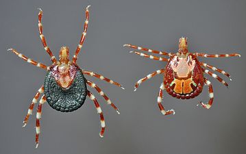 Amblyomma variegatum ixodid ticks, female and male, dorsal.
 -
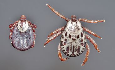 Dermacentor andersoni ixodid ticks, female and male, dorsal.
 -
 Haemaphysalis bancrofti ixodid ticks, female and male, dorsal.
 -
 Ixodes holocyclus ixodid ticks, female and male, dorsal.


