
Wikipedia:WikiProject AP Biology 2016

1. DNA
2. Enhancer
3. Promoter
4. Gene
5. Transcription Activator Protein
6. Mediator Protein
7. RNA Polymerase
- Past Related Projects: Wikipedia:WikiProject AP Biology Bapst 2012, Wikipedia:WikiProject AP Biology Bapst 2013, Wikipedia:WikiProject AP Biology Bapst 2014, & Wikipedia:WikiProject AP Biology Bapst 2015
A high school class in
- Students will work alone, there are 43 students so we should have 43 new images with captions and labels.
- The time frame will be three weeks.
- Students will be required to write a summary of why they select a topic; hopefully, eliminating obscure, random topic selections. They also must create labels and captions for their photos
- They may add it to encyclopedia articles.
- The best of the bunch will be submitted as here. Featured images must be in .svg (vector) format.
Feel free to discuss this project. Please notify me of any concerns; especially if they involve the behavior of my students on Wikipedia. With a little patience, this should be an inspirational experience for all.
Goals / Motivation
- To create a situation that not only vigorously enhances our ability to make quality decisions but also to improve our traction on the roads of 'Merica
- To improve the images in Wikipedia's coverage of Biology articles.
- To encourage promising students to write, create, learn, and contribute volunteer efforts through a service learningproject.
- The dreaded “Research Project” is a standard hurdle for most AP Programs. Rightfully so, being that many college courses require such publications to validate your existence. This new approach to constructing a scientific document, is far more authentic and interesting. Rather than researching for a paper that is destined for the teacher's eyes and then a one way trip to the circular bin, let us contribute to the world-wide data base for others to benefit. I hope this will be an interesting and memorable project and assessment. It's funny, I can remember a number of projects and papers I wrote during my own high school experience, but I can remember no tests whatsoever.
Contributions
As you upload your projects and add them to Wikipedia please add them to the gallery below. By adding a new line which begins with the word "File" and them follows the format of my sample image. Make sure to include your caption.
-
 Toxoplasmosis life cycle:
1.Cat eats prey with toxoplasmosis becoming the primary host of Toxoplasma gondii.
2. Toxoplasma gondii disrupts the wall of the cat’s small intestine, forming oocysts, as well as tissue cysts in the brain and muscles.
3.When the cat excretes waste, the feces is contaminated with the oocysts.
4. The feces contaminates the surrounding plants.
5. Organisms eat the plants consuming the oocysts, and become infected by the parasite. When consumed by another organism, the parasite invades the new host, causing the cycle to begin again. -
 This image represents different types of sequences following TATA boxes.The TATA boxes above represent the following sequences:
E.Coli a70 Promoter BBa_J010062
E. Coli a70 Promoter BBa_J231083
Yeast Promoter BBa_K122000 -
 The cyclic light-dependent reactions occur when only the sole photosystem being used is photosystem 1. Photosystem 1 excites electrons which then cycle from the transport protein, ferredoxin (Fd), to the cytochrome complex, b6f, to another transport protein, plastocyanin (Pc), and back to photosystem I. A proton gradient is created across the thylakoid membrane (6) as protons (3) are transported from the chloroplast stroma (4) to the thylakoid lumen (5). Through chemiosmosis, ATP (9) is produced where ATP synthase (1) binds an inorganic phosphate group (8) to an ADP molecule (7).
 -
 A neural pathway connects a part of the nervous system to another using bundles of axons. The optic nerve is an example of a neural pathway because it connects the eye back to the brain.
 -
 Excreted from the pancreas, insulin circulates through the blood before binding to an insulin receptor on a fat or muscle cell. Once insulin binds to the receptor, phosphorylation takes place and attaches to the beta-subunit, thus initiating the transduction process. A protein binds to the phosphorylated receptor protein, becoming phosphorylated as well. Once the protein detaches from the receptor protein, the signal has successfully been transported from the receptor to the newly active protein. Through a series of kinase proteins, the proteins are constantly being phosphorylated and activated. At the end of the transduction process, the activated protein binds to the PIP2 proteins embedded in the membrane. By doing so, the initial signal has successfully transmitted the extracellular signal. As a result, another protein is activated, later activating the storage vesicles found within the cell. Upon activation, the vesicle is transported to the membrane, where its membrane becomes integrated within the cellular membrane under a process known as phagocytosis. The Glut-4 protein channels that were once embedded in the storage vesicles are now embedded in the cellular membrane. Glucose can now flow into the cell through these glucose transport channels.
 -
![1. Tabular 2. Wedge 3. Dome 4. Drydock 5. Pinnacled 6. Blocky]]](//upload.wikimedia.org/wikipedia/commons/thumb/6/61/Types_of_Icebergs.svg/120px-Types_of_Icebergs.svg.png) 1. Tabular 2. Wedge 3. Dome 4. Drydock 5. Pinnacled 6. Blocky]]
1. Tabular 2. Wedge 3. Dome 4. Drydock 5. Pinnacled 6. Blocky]] -
 1. Epipelagic zone: surface - 200 meters deep 2. Mesopelagic zone: 200 - 1000m3. Bathypelagic zone: 1000m - 4000m 4. Abyssopelagic zone: 4000m - 6000m5. Hadal zone (the trenches): 6000 m to the bottom of the ocean
 -
![1. Sprout 2. Dormant bud 3. Periderm 4. Cortex 5. Vascular Ring 6. Perimedulla7. Outer medulla]]](//upload.wikimedia.org/wikipedia/commons/thumb/8/8c/BIO_EXTRA_CREDIT.svg/120px-BIO_EXTRA_CREDIT.svg.png) 1. Sprout 2. Dormant bud 3. Periderm 4. Cortex 5. Vascular Ring 6. Perimedulla7. Outer medulla]]
1. Sprout 2. Dormant bud 3. Periderm 4. Cortex 5. Vascular Ring 6. Perimedulla7. Outer medulla]] -
 Character displacement occurs when similar species that live in the same geographical region and occupy similar niches differentiate in order to minimize niche overlap and avoid competitive exclusion. Several species of Galapagos finches, shown above, display character displacement. Each closely-related species differs in beak size and beak depth, allowing them to coexist in the same region since each species eats a different type of seed: the seed best fit for its unique beak. The finches with the deeper, stronger beaks consume large, tough seeds, while the finches with smaller beaks consume the smaller, softer seeds.
 -
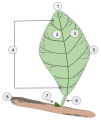 Diagram of a Simple Leaf1. Apex 2. Midvein (Primary vein)3. Secondary vein. 4. Lamina.5. Leaf margin6. Petiole7. Bud8. Stem
 -
 There are three different types of cartilage; hyaline (A), elastic (B), and fibrous (C). In elastic cartilage the cells are closer together creating less intercellular space. Elastic cartilage is found in the external ear flaps and in parts of the larynx. Hyaline cartilage has less cells than elastic cartilage, there is more intercellular space. Hyaline cartilage is found in the nose, ears, trachea, parts of the larynx, and smaller respiratory tubes. Fibrous cartilage has the least amount of cells so it has the most amount of intercellular space. Fibrous cartilage is found in the spine and the menisci.
 -
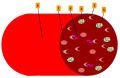 This vein (4) shows the interaction between the malaria sporozoites (6) with sickle cells (3) and regular cells (1). While malaria is still affecting the regular cells (2), the ratio of sickle to regular cells is 50/50 due to sickle cell anemia being a heterozygous trait, so the malaria can’t affect enough cells with schizonts (5) to harm the body.
 -
 A. Lancelet, B. Larval tunicate, C. Adult tunicate. 1. Notochord, 2. Nerve chord, 3. Buccal cirri, 4. Pharynx, 5. Gill slit, 6. Gonad, 7. Gut, 8. V-shaped muscles, 9. Anus, 10. Inhalant syphon, 11. Exhalant syphon, 12. Heart, 13. Stomach, 14. Esophagus, 15. Intestines, 16. Tail, 17. Atrium, 18. Tunic.
 -
![There are three basic variants of immunoglobulin antigens in humans that share a very similar chemical structure but are distinctly different. Red circles show where there are differences in chemical structure in the antigen-binding site (sometimes called the antibody-combining site) of human immunoglobulin. Notice the O-type antigen does not have a binding site.[1]](//upload.wikimedia.org/wikipedia/commons/thumb/a/aa/AP-Biology_Final_Project.svg/88px-AP-Biology_Final_Project.svg.png) There are three basic variants of immunoglobulin antigens in humans that share a very similar chemical structure but are distinctly different. Red circles show where there are differences in chemical structure in the antigen-binding site (sometimes called the antibody-combining site) of human immunoglobulin. Notice the O-type antigen does not have a binding site.[1]
There are three basic variants of immunoglobulin antigens in humans that share a very similar chemical structure but are distinctly different. Red circles show where there are differences in chemical structure in the antigen-binding site (sometimes called the antibody-combining site) of human immunoglobulin. Notice the O-type antigen does not have a binding site.[1] -
 The dinoflagellate labeled above is the microscopic algae Karenia brevis. Karenia brevis is the cause of red tide in the Gulf of Mexico. The algae propels itself by using a longitudinal flagellum (A) and a transverse flagellum (B). The longitudinal flagellum lies in a groove- like structure called the cingulum (F). The dinoflagellate is separated into an upper portion called the epitheca (C) where the apical horn resides (E) and a lower portion called the hypotheca (D). Photo used from https://en.wikipedia.org/wiki/Karenia_brevis#/media/File:Karenia_brevis.jpg
 -
![The reproduction mechanism of a typical viroid. Leaf contact transmits the viroid. The viroid enters the cell via its plasmodesmata. RNA polymerase II catalyzes rolling-circle synthesis of new viroids.]]](//upload.wikimedia.org/wikipedia/commons/thumb/0/05/Viroids-_how_it_do.gif/120px-Viroids-_how_it_do.gif) The reproduction mechanism of a typical viroid. Leaf contact transmits the viroid. The viroid enters the cell via its plasmodesmata. RNA polymerase II catalyzes rolling-circle synthesis of new viroids.]]
The reproduction mechanism of a typical viroid. Leaf contact transmits the viroid. The viroid enters the cell via its plasmodesmata. RNA polymerase II catalyzes rolling-circle synthesis of new viroids.]] -
 Muscles will contract or relax when they receive signals from the nervous system. The neuromuscular junction is the site of the signal exchange. The steps of this process in vertebrates occur as follows:(`) The action potential reaches the axon terminal. (2) Voltage-dependent calcium gates open, allowing calcium to enter the axon terminal. (3) Neurotransmitter vesicles fuse with the presynaptic membrane and acetylcholine (ACh) is released into the synaptic cleft via exocytosis. (4) ACh binds to postsynaptic receptors on the sarcolemma. (5) This binding causes ion channels to open and allows sodium ions to flow across the membrane into the muscle cell. (6) The flow of sodium ions across the membrane into the muscle cell generates an action potential which travels to the myofibril and results in muscle contraction. Labels: A: Motor Neuron Axon B: Axon Terminal C: Synaptic Cleft D: Muscle Cell E: Part of a Myofibril
 -
 The red shading indicates the range of the bonobo. The blue shading indicates the range of the chimpanzee. This is an example of allopatric speciation because they are divided by a natural barrier (the Congo River) and have no habitat in common. / L'ombre rouge signifie ou habitent les bonobos. L'ombre bleu signifie ou habitent les chimpanzés. C'est une example de spéciation allopatrique parce qu'ils sont séparés par une barrière naturale (le rivière Congo) et ne partage rien du habitat.
 -
 The red shading indicates the range of the brown bear. The blue shading indicates the range of the polar bear. The purple shading indicates areas where both live. This is an example of peripatric speciation because polar bears' ancestors moved to a new ecological niche on the periphery of the brown bears' original habitat. / L'ombre rouge signifie ou habitent les ours bruns. L'ombre bleu signifie ou habitent les ours polaires. L'ombre violet signifie les lieux ou habitent les deux espèces des ours. C'est une example de spéciation péripatrique parce que les ancestres des ours polaires se sont bougés à un nouveau niche écologique sur le bord du habitat original des ours bruns.
 -
 The spleen contains two different tissues, white pulp (A) and red pulp (B). The white pulp functions in producing and growing immune and blood cells. The red pulp functions in filtering blood of antigens, microorganisms, and defective or worn-out red blood cells.
 -
 Classifications of Chromosomes
I: Telocentric - centromere placement very close to the top, p arms barely visible if visible at all
II: Acrocentric - q arms are still much longer than the p arms, but the p arms are longer than it those in telocentric
III: Submetacentric - p and q arms are very close in length but not equal
IV: Metacentric - the p arm and the q arms are equal in length
A: Short arm (p arm)
B: Centromere
C: Long arm (q arm)
D: Sister Chromatid -
 The C4 pathway contains a special structure in the leaves known as Kranz anatomy. There exists a layer of mesophyll cells that contain small chloroplasts encircling bundle sheath cells that have large chloroplasts necessary for the Calvin cycle.
A: Mesophyll Cell
B: Chloroplast
C: Vascular Tissue
D: Bundle Sheath Cell
E: Stroma
F: Vascular Tissue: provides continuous source of water
1) Carbon is fixed to produce oxaloacetate by PEP carboxylase.
2) The four carbon molecule then exits the cell and enters the chloroplasts of bundle sheath cells.
3) It is then broken down releasing carbon dioxide and producing pyruvate. Carbon dioxide combines with ribulose bisphosphate and proceeds to the Calvin Cycle.
4) Pyruvate re-enters the mesophyll cell. It then reacts with ATP to produce the beginning compound of the C4 cycle. -
 The BRCA genes are tumor suppressor genes pictured here on their respective chromosomes. BRCA 1 has the cytogenetic location 17q21 or the q arm of Chromosome 17 at position 21. BRCA 2 has the cytogenetic location 13q12.3 or the q arm of Chromosome 13 at position 12.3. Both genes produce proteins that help repair damaged DNA, keeping the genetic material of the cell stable. A damaged BRCA gene in either location can lead to increased risk of cancer, particularly breast or ovarian in women.
 -
![Reindeer introduced to St. Matthew Island in 1944 increased from 29 animals at that time to 6,000 in the summer of 1963, a drastic overshoot of the island’s carrying capacity causing a crash die-off the following winter to 42 animals. Based on the size of the island, recent estimates put the carrying capacity at about 1,670 animals [Klein, D. R. (n.d.). The Introduction, Increase, and Crash of Reindeer on St. Matthew Island. Retrieved May 25, 2016, from http://dieoff.org/page80.htm].](//upload.wikimedia.org/wikipedia/commons/thumb/7/7a/St._Matthew_Island_Reindeer_Population.svg/120px-St._Matthew_Island_Reindeer_Population.svg.png) Reindeer introduced to St. Matthew Island in 1944 increased from 29 animals at that time to 6,000 in the summer of 1963, a drastic overshoot of the island’s carrying capacity causing a crash die-off the following winter to 42 animals. Based on the size of the island, recent estimates put the carrying capacity at about 1,670 animals [Klein, D. R. (n.d.). The Introduction, Increase, and Crash of Reindeer on St. Matthew Island. Retrieved May 25, 2016, from http://dieoff.org/page80.htm].
Reindeer introduced to St. Matthew Island in 1944 increased from 29 animals at that time to 6,000 in the summer of 1963, a drastic overshoot of the island’s carrying capacity causing a crash die-off the following winter to 42 animals. Based on the size of the island, recent estimates put the carrying capacity at about 1,670 animals [Klein, D. R. (n.d.). The Introduction, Increase, and Crash of Reindeer on St. Matthew Island. Retrieved May 25, 2016, from http://dieoff.org/page80.htm]. -
 In both stages of metamorphosis, the insect begins the cycle as an egg. In a complete metamorphosis the insect passes through four distinct phases which produce an adult that does not resemble the larvae. In an incomplete metamorphosis an insect does not go through a full transformation, but instead transitions from a nymph to an adult by molting its exoskeleton whenever it becomes too tight.https://www.cals.ncsu.edu/course/ent425/tutorial/morphogenesis.html
 -
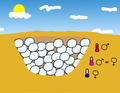 Some reptiles use incubation temperatures to determine their sex. In some species this follows the pattern that eggs in high temperatures become male and eggs that develop in cooler temperatures become female.
 -
 Some reptiles use incubation temperatures to determine sex. In some species this follows the pattern that eggs in extreme high or low temperatures become male and eggs in medium temperatures become female.
 -
 Hemoglobin and Chlorophyll, two extremely different molecules when it comes to function, are amazingly similar when it comes to its atomic shape.There are only three major structural differences; a magnesium atom (Mg) in chlorophyll, which is replaced with iron (Fe) in hemoglobin. Additionally, chlorophyll has some extra structures on the bottom right side (A), and an extended hydrocarbon tail on the left (B). These differences cause the chlorophyll molecule to be non-polar, in contrast to the polar hemoglobin molecule.
 -
 Graph shows principles of Intermediate Disturbance Hypothesis: I. at low levels of ecological disturbance species richness decreases as competitive exclusion increases, II. at intermediate levels of disturbance, diversity is maximized because species that thrive at both early and late successional stages can coexist, III. at high levels of disturbance species richness is decreased due an increase in species movement.
 -
 Enzyme inhibitors slow or stop the catalytic activity of enzymes by binding with their active sites.(A) Competitive inhibitors interfere directly with an enzyme’s binding site to ensure the substrate cannot fit. (B) Non-competitive inhibitors bind to an allosteric site of an enzyme, changing its shape
 -
 Cladogram of the family Balaenopteridae using complete mtDNA sequences and short interspersed repetitive element (SINE) insertion data. Mysticeti. Cetacean Palaeobiology. N.p., n.d. Web. 31 May 2016.
 -
 Mammals and insects are part of different homologous and analogous evolutionary groups. In the horizontal direction, the structures are homologous in their morphology, or anatomy, but different in their function due to differences in habitat. In the vertical direction, the structures are analogous in function due to similar lifestyles of organisms but anatomically different since they are part of different groups.
 -
 Sexual selection is a form of natural selection where one sex prefers a specific characteristic in an individual of the other sex. Peafowls exhibit sexual selection in that peahens look for peacocks who have more “eyes” on their tail feathers. If a peacock has fewer “eyes”, then the peahen will continue to look for a better, more suitable mate. This will eventually cause the peacocks with fewer eyes to die out and the peacocks with more “eyes” to continue to grow in proportion to the population size.
 -
 Often used as a cancer treatment in postmenopausal women, aromatase inhibitors work by blocking the conversion of androstenedione and testosterone into estrone and estradiol, respectively, which are both crucial to the growth of developing breast cancers (aromatase inhibitors are also effective at treating ovarian cancer, but less commonly so). In the diagram, the adrenal gland (1) releases androstenedione (3) while the ovaries (2) secrete testosterone (4). Both hormones travel to peripheral tissues or a breast cell (5), where they would be converted into estrone (8) or estradiol (9) if not for aromatase inhibitors (7), which prevent the enzyme CYP19AI (also known as aromatase or estrogen synthase) (6) from catalyzing the reaction that turns androstenedione and testosterone into estrone and estradiol. In the diagram, Part A represents the successful conversion of androstenedione and testosterone into estrone and estradiol in the liver. Part B represents the blockage of this conversion by aromatase inhibitors both in peripheral tissues and in the breast tumor itself.
 -
 Blood glucose levels are maintained at a constant level in the body by a negative feedback mechanism. When the blood glucose level is too high, the pancreas secretes insulin and when the level is too low, the pancreas then secretes glucagon. The flat line shown represents the homeostatic set point. The sinusoidal line represents the blood glucose level.
 -
 Lamarckian inheritance is the idea that individuals can pass down traits acquired during their lifetime to their offspring. This diagram is an example that supports Lamarckian inheritance. The first giraffe has a short neck and cannot reach the leaves to eat. Over time the first giraffe would create a gene that would say it needed a longer neck to reach the food and it would then pass down the trait to its offspring to have a longer neck, so it could reach the food. By the fourth giraffe, it would be able to reach the leaves due to its extended neck length, which has changed over multiple generations.
 -
 This animation focuses on one molecule of glucose turning into pyruvate then into lactic acid.
 -
 In 1966 Microbiologist Kwang Jeon conducted an experiment with amoebae communities providing real-life evidence for the endosymbiotic theory.
 -
 Ribonucleoproteins (snRNPs) are subunits of spliceosomes (small RNA and proteins). SnRNPs cut the DNA and remove noncoding introns (2) from the coding exons (1). This is an accessible process to producing a mature mRNA for RNA translation. The introns are cut at the 5’ and 3’ splice sites.The U1 snRNP binds to the 5’ splice site, while U6 snRNP binds to the 3’ splice site. Then U2, U4, U5 binds in the intron region (branch site). The 5’ splice site on the end of the intron is cut first. The free 5’ end then bonded to an adenine in the branch site.U1 and U4 snRNPs are released as the U3, U5, U6 snRNPs shift on the lariat (the removed intron with the snRNPs attached). The 3’ splice site is cut and the exon sites are connected. The lariat then disintegrates away from the exons.Mature mRNA is left.
 -
 1.Haploid organisms are organisms which have only one set of chromosomes, they are on the left. Diploid organism are organisms which have two sets of chromosomes, they are on the right. 2.In this model, purple individuals express the dominant mendelian genes. This is an example of a egg from a haploid individual carrying the dominant genes.3.In this model, blue organisms express the recessive mendelian genes. This is an example of a sperm from a haploid individual carrying the recessive genes.4.This is a sperm from a diploid individual that is carrying a recessive gene for a blue color.5.This is a egg from a diploid individual that is carrying a dominant gene for a purple color.6.In a Haploid life cycle (left) for a short time they have a diploid structure so they can produce spores through meiosis7.This is the first stage of a zygote which has just been fertilized by a sperm.8.The spores released by the diploid structure either express the mothers dominate gene or the fathers recessive gene. 9.With the cells of the baby, everyone express the dominant gene but has the recessive genes.
 -
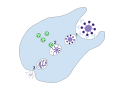
 When a cell is infected, the helper T Cells direct the cytotoxic T Cells to destroy the cell. Using small HMC’s located all over the cell which contain fragments of a pathogen, the infected cell is detected and latched onto by the T Cell. Perforin breaks the membrane and the lymphotoxin flows in and kills the cell.1) One cell is infected by a virus. The Histocompatibility Complexes located all around the cell have parts of the virus on them, allowing the T Cell to detect the infected cell.2) A) Pathogen B) T Cell Receptor Complex (TCR) C) T Cell D) Lymphotoxin E) Lymphokine F) Major Histocompatibility Complex (MHC)3) G) Perforin H) CD8 4) Antigen is destroyed and cell components flow out through the now porous membrane.Ignore changes in cell size. MHCs are located all over the cell. One was enlarged for simplicity in this diagram.
 -
 1. Excess nutrients are applied to the soil. 2. Some nutrients leach into the soil where they can remain for years. Eventually, they get drained into the water body. 3. Some nutrients run off over the ground into the body of water. 4. The excess nutrients cause an algal bloom. 5. The algal bloom blocks the light of the sun from reaching the bottom of the water body. 6. The plants beneath the algal bloom die because they cannot get sunlight to photosynthesize. 7. Eventually, the algal bloom dies and sinks to the bottom of the lake. Bacteria begins to decompose the remains, using up oxygen for respiration. 8. The decomposition causes the water to become depleted of oxygen. Larger life forms, such as fish, suffocate to death. This body of water can no longer support life.
 -
 Phagocytosis is the process in which a cell engulfs a particle, digests it, and expels the waste products. Process of phagocytosis: 1. A particle is ingested by a phagocyte after antigens are recognized which results in the formation of a phagosome. 2. The fusion of lysosomes with the phagosome creates a phagolysosome. 3. The particle is broken down by the digestive enzymes found in the lysosomes. The resulting waste material is discharged from the phagocyte by exocytosis.
 -
 Pre-mRNA is the first form of RNA created through transcription in protein synthesis. The pre-mRNA lacks structures that the messenger RNA (mRNA) requires. First all introns have to be removed from the transcribed RNA through a process known as splicing. Before the RNA is ready for export, a Poly(A)tail is added to the 3’ end of the RNA and a 5’ cap is added to the 5’ end.
 -
 The ligands, denoted by letter L, signal for platelets (P) to migrate towards the wound (Site A). As more platelets gather around the opening, they produce more ligands to amplify the response. The platelets congregate around the wound in order to create a cap to stop blood flow out of the tissue.
 -
 Environmental DNA (eDNA) is DNA that an organism leaves behind as it moves through an environment. Generally, eDNA follows a pattern of exponential decay over time. The fish above leaves its eDNA behind as it moves through the aquatic environment. The eDNA stays behind in the environment, and slowly dissipates over time. Barnes, M. A., Turner, C. R., Jerde, C. L., Renshaw, M. A., Chadderton, W. L., & Lodge, D. M. (2014). Environmental Conditions Influence eDNA Persistence in Aquatic Systems. Environmental Science & Technology Environ. Sci. Technol., 48(3), 1819-1827. doi:10.1021/es404734p
 -
 In a reflex arc, an action potential never travels to the brain for processing and so results in a much quicker reaction. When a stimulus (A) is encountered, the signal from that stimulus will travel up the sensory neuron (B, in green) to the spinal column (C). There, it will likely pass through a short interneuron (D, in purple) before continuing down a motor neuron (E, in blue) to the origin of the signal. Then, a contraction of the muscles (F) is triggered, moving the bone (G).
 -
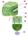
 A single diatom at first look may not seem to be a cell, but diatoms are eukaryotic organisms with common organelles such as a nucleus, mitochondria, and golgi complex.
1) Nucleus; holds the genetic material
2) Nucleolus; Location of the chromosomes
3) Golgi complex; modifies proteins and sends them out of the cell
4) Cell Wall; Outer membrane of the cell5) Pyrenoid; center of carbon fixation
6) Chromatophore; pigment carrying membrane structure
7) Vacuoles; vesicle of a cell that contains fluid bound by a membrane8) Cytoplasmic strands; hold the nucleus
9) Mitochondria; creates ATP (energy) for the cell
10) Valve/Striae; allows nutrients and waste in and out of the cell
.svg)


![1. Tabular 2. Wedge 3. Dome 4. Drydock 5. Pinnacled 6. Blocky]]](/File:Types_of_Icebergs.svg)
.svg)
![1. Sprout 2. Dormant bud 3. Periderm 4. Cortex 5. Vascular Ring 6. Perimedulla7. Outer medulla]]](/File:BIO_EXTRA_CREDIT.svg)


![There are three basic variants of immunoglobulin antigens in humans that share a very similar chemical structure but are distinctly different. Red circles show where there are differences in chemical structure in the antigen-binding site (sometimes called the antibody-combining site) of human immunoglobulin. Notice the O-type antigen does not have a binding site.[1]](/File:AP-Biology_Final_Project.svg)
.png)
![The reproduction mechanism of a typical viroid. Leaf contact transmits the viroid. The viroid enters the cell via its plasmodesmata. RNA polymerase II catalyzes rolling-circle synthesis of new viroids.]]](/File:Viroids-_how_it_do.gif)


![Reindeer introduced to St. Matthew Island in 1944 increased from 29 animals at that time to 6,000 in the summer of 1963, a drastic overshoot of the island’s carrying capacity causing a crash die-off the following winter to 42 animals. Based on the size of the island, recent estimates put the carrying capacity at about 1,670 animals [Klein, D. R. (n.d.). The Introduction, Increase, and Crash of Reindeer on St. Matthew Island. Retrieved May 25, 2016, from http://dieoff.org/page80.htm].](/File:St._Matthew_Island_Reindeer_Population.svg)


.gif)


Contributors
Add your user name here following my example. Just add this template with your username instead of the line: {{user|username}} and then, if your username is not identifiable, your real first name.
- Earthdirt (talk · contribs) - Chris (AKA Mr. Packard) - IMAGE TOPIC NAME HERE
- apawnilation (talk · contribs) - David - TATA Box
- Verona Dethran (talk · contribs) - Brendan - Reflex Arc
- Bbowen23 (talk · contribs) - Brooke - Positive Feedback Loop
- Kungfucrab (talk · contribs) - Sarah - Eutrophication
- planetseeker (talk · contribs) - MaggieBeth - Temperature-dependent sex determination
- Marabouchoklad (talk · contribs) - Meredith - Endosymbiotic Theory- 2004 Jeon experiment
- helloportobello (talk · contribs) - Natalie - Negative Feedback Loop
- Senioritisisreal (talk · contribs) - Danyel - Spleen
- I Am Not Original6 (talk · contribs) - Susannah - Lactic Acid Fermentation
- Modojo (talk · contribs) - Morgan- snRNP
- sizzlinggreg (talk · contribs) - Gina - Sexual Selection
- Mango Slices (talk · contribs) - Julia - Phagocytosis
- meimeit21 (talk · contribs) - Mei - Cyclic Light Dependent Reactions
- Yeswikipediaimarobot (talk · contribs) - Victoria - eDNA
- Basketball1713 (talk · contribs) - Lauren - Tunicate & Sea Squirt
- universalwin1222 (talk · contribs) - Emma - Lamarckian inheritance
- Jcauctkting (talk · contribs) - Jack - Hemoglobin/chlorophyll
- Mads00 (talk · contribs) - Maddie - Red Queen Hypothesis
- Ilovesquirrelz (talk · contribs) - Lily - Aromatase inhibitors
- AlexBatchelder (talk · contribs) - Ben - Malaria and Sickle Cell Anemia
- Asychterz18 (talk · contribs) - Aleksei - Haploid Genetics
- liquiddeer (talk · contribs) - Isaac - Viroids
- Clumsybatman (talk · contribs) - Elyse - Diatom
- spencerAC (talk · contribs) - Spencer - Killer T Cells
- Fockey003 (talk · contribs) - Kaylah - Centromeres and positioning
- Awakening Conscience (talk · contribs) - Clara - Character Displacement
- adamruns (talk · contribs) - Adam - C4 Plants
- OutletPlug (talk · contribs) - Brandon - Molecular Clock
- yuppo1 (talk · contribs) - Bryson - Alcoholic Fermentation
- Username1927 (talk · contribs) - Haley - Holometabolism vs. Hemimetabolism
- tessssa13 (talk · contribs)- Tessa- BRCA mutation
- Ihadpieforlunch (talk · contribs) - Gage - Blood transfusion
- Luuis12321 (talk · contribs) - Vicky - Signal Transduction Insulin
- california16 (talk · contribs)- callie - Competitive/Noncompetitive Inhibition
- Shiloh117981894 (talk · contribs)- Rachel - Cartilage
- CPiGuy (talk · contribs) - Conor - Peripatric Speciation
- nastypatty (talk · contribs) - Alexander Batchelder - lncRNA
- ratchelp11 (talk · contribs) - Rachel P. - Red tide and types of algal blooms
- MattyGprotein (talk · contribs) - Matt Greenlaw - Rorquals Cladogram
- sciencerelatedusername (talk · contribs) - Elizabeth - Intermediate Disturbance Hypothesis
- vanessablakegraham (talk · contribs) - Vanessa Blake Graham - Convergent Evolution
- ilovericexoxo (talk · contribs) - Aylee Doane - 'Toxoplasmosis
Uploading
In order to complete the assignment and reap all the benefits of your hard work (such as a good grade) you MUST complete all of the following steps. If you need help, just ask.
How to, step by step
Step 1: Create a Wikipedia Global account by clicking "Login/create account" in the upper right hand corner of this page.
Step 2: Click here to use the WikiCommons File Upload Wizard
Step 3: If you didn't do it in the Wizard, categorize your image by adding a one or more [[Category:_______]] tags at the bottom of the page (fill in the name of the category in the _______.) You might use Category:Biology diagrams (but that's not a very helpful category) or something more specific like Category:Molecular biology or something else appropriate.
Step 4: If you didn't do it in the Wizard you should also now add your labels and your caption information in the description to your upload page in the Commons.
Step 5: Your image is now available in all Wiki Projects, including Wikipedia. So let's add it to the article! Go to the article you want to add your donated image to. In the top of the section of the article or the subheading you want to add the image to add something like this:
[[File:MY IMAGE NAME.png|right|thumb|200px|The [[caption]] of '''my image'''.]]
That's not too hard is it? For your caption you'll need to follow Wikipedia style and use some mark up to do this - it's kind of like a micro-essay. The [[ ]] creates a link to the given page on Wikipedia and the ''' ''' make the word bold, in Wikipedia it's appropriate to bold the title of the article the first time it's used in the text or in a caption."
Step 6: Wow you've done it! Now you just have to turn in your work by adding it to gallery in the section above here called "Contributions". Just follow the model I provided in the first entry. Make sure that your entry is between the <gallery> and </gallery> tags or it won't show up. Your caption will likely have to be shorter than your description, see the style advice below.
Style guides
To get past the stumbling blocks of editing Wikipedia, articles will have to conform to the Wikipedia style guides. The largest barriers are:
- Wikipedia:Picture tutorialis also useful.
- Wikipedia:Manual of Style/Captions - Writing a good caption may be harder than you think.
- Wikipedia:Copyrights - Make sure to post a license on your image which releases all copyrights and makes it free use image AND don't use images from anywhere except the Commons if your image integrates other images.
- Wikipedia:File names - Pick the right name for your file.
- Wikipedia:Preparing images for upload - Pick the right file type (images created using entirely Google Draw should be saved as .SVG, whereas most other images you make will be saved as a .PNG in rare cases an a .JPG or .JPEG can be used)
- Wikipedia:Uploading images or WikiCommons Uploading Images - Do it right the first time (or just use the Wizard).
- Wikipedia:Ten things you may not know about images on Wikipedia - Kind of interesting.
You can always ask for help at:
- The Help Deskor
Writing a good image caption
There are several criteria for a good caption. A good caption:
- clearly identifies the subject of the picture, without detailing the obvious.
- is succinct (that means short).
- establishes the picture's relevance to the article.
- provides context for the picture.
- draws the reader into the article.
Different people read articles different ways. Some people start at the top and read each word until the end. Others read the first paragraph and scan through for other interesting information, looking especially at pictures and captions. For those readers, even if the information is adjacent in the text, they will not find it unless it is in the caption—but do not tell the whole story in the caption—use the caption to make the reader curious about the subject.
Another way of approaching the job: imagine you're giving a lecture based on the encyclopedia article, and you are using the image to illustrate the lecture. What would you say while attention is on the image? What do you want your audience to notice in the image, and why? Corollary: if you have got nothing to say, then the image probably does not belong in the article.
Images for the lead
It is very common to use an appropriate representative image for the lead of an article, often as part of an infobox. The image helps to provide a visual association for the topic, and allows readers to quickly assess if they have arrived at the right page. For most topics, the selection of a lead image is plainly obvious: a photograph or artistic work of a person, photographs of a city, or a cover of a book or album, to name a few.
Image selection for other topics may be more difficult and several possible choices could be made. While Wikipedia is not censored, as outlined in the above section on offensive images, the selection of the lead image should be made with some care with respect to this advice. Lead images are loaded and shown upon navigating to the page, and are one of the first things that readers will see. Editors should avoid using images that readers would not have expected to see when navigating to the page. Unlike other content on a page that falls below the lead, the lead image should be chosen with these considerations in mind.
Some advice on selecting a lead image include the following:
- Lead images should be images that are natural and appropriate visual representations of the topic; they not only should be illustrating the topic specifically, but should also be the type of image that is used for similar purposes in high-quality reference works, and therefore what our readers will expect to see. Lead images are not required, and not having a lead image may be the best solution if there is no easy representation of the topic.
- Lead images should be selected to be of least shock value; if an alternative image exists that still is an accurate representation of the topic but without shock value, it should always be preferred. For example, using an image of deportees being subjected to selection as the lead image at this version of Holocaustis far preferable to the appropriate images that appear later in the article that show the treatment of the prisoners or corpses from the camps.
- Sometimes it is impossible to avoid the use of a lead image with perceived shock value if the topic itself is of that nature, for example in articles on various parts of human genitalia. It should be anticipated, through Wikipedia:Content disclaimer, that readers will be aware they will be exposed to potentially shocking images when navigating to articles on such topics.
Planning and resources
- Wikipedia tutorials for beginners
- Editing commands cheatsheet
- Getting started
- The perfect article
- Assessment
- Article development
- Peer Review
- [[Active gif creator]]
Talk pages
These are places where you can leave and receive messages and questions, every page has one. Whenever you edit these pages, make sure that you are signed in. Also, add four tildes ~~~~ to the end of all comments you make on talk pages. This will let people know who is talking.
- PMID 24384923.