
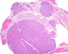
Canalicular adenoma
Canalicular adenoma is a type of growth that occurs in human salivary glands. It is a benign growth which occurs in the epithelial cells, and is typically arranged in columns of cells that form interconnecting cords. Canalicular adenoma is a very rare benign neoplasm; it constitutes about 1% of all salivary gland tumors and about 4% of all benign salivary gland tumors.[1][2]
Presentation
Canalicular adenoma is most common in patients age 70 to 80, with females affected about four times as often as males. Most growths present in the upper lip; some also occur in the a few present in palate or buccal (cheek) tissue as a slowly enlarging mass.[3] The growths will often arise in multiple places at the same time or develop multiple nodes, despite not being clinically invasive or malignant.[1][4]
Diagnosis
Canalicular adenoma growths are usually small at the time they are noticed, with an average size of about 1.6 cm.
Treatment
Most instances of canalicular adenoma are treated with conservative surgery.[1]