
Dermatome (anatomy)
| Dermatome | |
|---|---|
 Dermatomes of the upper and lower limbs (modified, after Keegan, J. J., and Garrett, F. D.) | |
 Dermatomes of the upper parts of the body, displaying significant overlapping (modified, from Fender, after Foerster) | |
| Anatomical terminology |
A dermatome is an area of
There are 8The term is also used to refer to a part of an embryonic somite.
Along the thorax and abdomen, the dermatomes are like a stack of discs forming a human, each supplied by a different spinal nerve. Along the arms and the legs, the pattern is different: the dermatomes run longitudinally along the limbs. Although the general pattern is similar in all people, the precise areas of innervation are as unique to an individual as fingerprints.
An area of skin innervated by a single nerve is called a peripheral nerve field.
The word dermatome is formed from Ancient Greek δέρμα 'skin, hide' and τέμνω 'cut'.
Clinical significance

A dermatome is an area of skin supplied by sensory neurons that arise from a spinal nerve ganglion. Symptoms that follow a dermatome (e.g. like pain or a rash) may indicate a pathology that involves the related nerve root. Examples include somatic dysfunction of the spine or viral infection. Certain skin problems tend to orient the lesions in the dermatomal direction.
In
Viruses that lie dormant in nerve ganglia (e.g. varicella zoster virus, which causes both chickenpox and shingles), often cause either pain, rash or both in a pattern defined by a dermatome (a zosteriform pattern). However, the symptoms may not appear across the entire dermatome.
Important dermatomes and anatomical landmarks
Following is a list of spinal nerves and points that are characteristically belonging to the dermatome of each nerve:[4]

- base of the skull. Alternately, a point at least 3 cm (1.2 in) behind the ear.
- midclavicular line.
- C4 – Over the acromioclavicular joint.
- antecubital fossa, just proximally to the elbow.
- proximal phalanx of the thumb.
- proximal phalanx of the middle finger.
- C8 – On the dorsal surface of the proximal phalanx of the little finger.
- antecubital fossa, just distal to the medial epicondyle of the humerus.
- T2 – At the apex of the axilla.
- midclavicular line and the third intercostal space
- midclavicular lineand the fourth intercostal space, located at the level of the nipples.
- T5 – Intersection of the midclavicular line and the fifth intercostal space, horizontally located midway between the level of the nipples and the level of the xiphoid process.
- T6 – Intersection of the midclavicular line and the horizontal level of the xiphoid process.
- T7 – Intersection of the midclavicular line and the horizontal level at one quarter the distance between the level of the xiphoid process and the level of the umbilicus.
- T8 – Intersection of the midclavicular line and the horizontal level at one half the distance between the level of the xiphoid process and the level of the umbilicus.
- T9 – Intersection of the midclavicular line and the horizontal level at three quarters of the distance between the level of the xiphoid process and the level of the umbilicus.
- T10 – Intersection of the midclavicular line, at the horizontal level of the umbilicus.
- T11 – Intersection of the midclavicular line, at the horizontal level midway between the level of the umbilicus and the inguinal ligament.
- T12 – Intersection of the midclavicular line and the midpoint of the inguinal ligament.
- L1 – Midway between the key sensory points for T12 and L2.[clarification needed]
- L2 – On the anterior medial thigh, at the midpoint of a line connecting the midpoint of the inguinal ligament and the medial epicondyle of the femur.
- L3 – At the medial epicondyle of the femur.
- medial malleolus.
- metatarsophalangeal joint.
- S1 – On the lateral aspect of the calcaneus.
- S2 – At the midpoint of the popliteal fossa.
- tuberosity of the ischium or intragluteal fold
- mucocutaneous zone
Following is a list of cranial nerves responsible for sensation from the face:
- V1 (1st division of the trigeminal nerve) - associated with herpes zoster ophthalmicus
- V2 (2nd division of the trigeminal nerve)
- V3 (3rd division of the trigeminal nerve)
Additional images
-
 Diagram of segmental distribution of the cutaneous nerves of the right upper extremity
 -
 Lower limb
 -
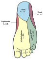 Foot
Foot -
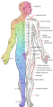 Major dermatomes and cutaneous nerves (anterior view)
 -
 Major dermatomes and cutaneous nerves (posterior view)


See also
- Cutaneous innervation
- Dorsal root
- Peripheral nerve field
References
- ^ Kishner, Stephen. "Dermatomes Anatomy". eMedicine. Medscape. Retrieved 2013-10-09.
- ^ "dermatome". The Free Dictionary by Farlex, Medical dictionary. Archived from the original on 2017-09-16.
- ^ "Referred Pain". Physiopedia. 2019-02-02. Archived from the original on 2019-05-21. cited van Cranenburghauthors, B. (1997). SCHEMA'S FYSIOLOGIE. Maarssen: Elsevier/De Tijdstroom. pp. 53, 65, 70.
- ^ "International Standards for the Classification of Spinal Cord Injury - Key Sensory Points" (PDF). American Spinal Injury Association. June 2008. Archived from the original (PDF) on 2016-03-04.
External links
- 3D Dermatomes Web App, Instamedic
- Hand kinesiology at the University of Kansas Medical Center
- Diagram of "Adult Dermatome", The New York Times