
Gallbladder polyp
| Gallbladder polyp | |
|---|---|
 | |
| A polyp in the gall bladder as seen on ultrasound |
Gallbladder polyps are growths or lesions resembling growths (polypoid lesions) in the wall of the gallbladder. True polyps are abnormal accumulations of mucous membrane tissue that would normally be shed by the body.
Signs and symptoms
Most polyps do not cause noticeable symptoms. Gallbladder polyps are usually found incidentally when examining the abdomen by ultrasound for other conditions, usually abdominal pain.[citation needed]
Pathology
Most small polyps (less than 1 cm) are not cancerous and may remain unchanged for years.
Cholesterolosis is characterized by an outgrowth of the mucosal lining of the gallbladder into fingerlike projections due to the excessive accumulation of
Adenomyomatosis describes a diseased state of the gallbladder in which the gallbladder wall is excessively thick, due to proliferation of subsurface cellular layer. It is characterized by deep folds into the
Diagnosis
Diagnosis is typically by ultrasound or CT imaging.
Upon histopathology of resected gallbladders, gallbladder polyps may be classified into the following main types:[4]
- Non-neoplastic polyps: Cholesterol, hyperplastic, and inflammatory polyps, adenomyomas, leiomyomas, fibromas, and lipomas
- Neoplastic polyps: adenomas, adenocarcinomas, and squamous cell carcinomas
-
 Ultrasound image of gallbladder polyps measuring 3–7 mm.
 -
![Gallbladder polyp types by relative incidence.[5]](//upload.wikimedia.org/wikipedia/commons/thumb/a/ac/Gallbladder_polyp_types_by_relative_incidence.jpg/460px-Gallbladder_polyp_types_by_relative_incidence.jpg) Gallbladder polyp types by relative incidence.[5]
Gallbladder polyp types by relative incidence.[5] -
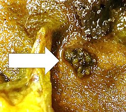 Photograph of a 6 mm large hyperplastic polyp of the gallbladder.
 -
 Histopathology of a hyperplastic polyp: There is no dysplasia.

![Gallbladder polyp types by relative incidence.[5]](/File:Gallbladder_polyp_types_by_relative_incidence.jpg)


Treatment
Most polyps are benign and do not need to be removed. Surgical removal of the gallbladder (
Epidemiology
Polypoid lesions of the gallbladder affect approximately 5% of the adult population.
The incidence of gallbladder polyps is higher among men than women. The overall prevalence among men of Chinese ancestry is 9.5%, higher than other ethnic types.[7]