
Lacunar ligament
| Lacunar ligament | |
|---|---|
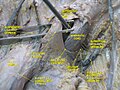
 The inguinal and lacunar ligaments. (Lacunar ligament labeled at center top.) | |
| Details | |
| From | inguinal ligament, pubic tubercle |
| To | pectineal line |
| Identifiers | |
| Latin | ligamentum lacunare (Gimbernati) |
| TA98 | A04.5.01.010 |
| TA2 | 2366 |
| FMA | 20184 |
| Anatomical terminology | |
The lacunar ligament, also named Gimbernat's ligament, is a ligament in the inguinal region.[1] It connects the inguinal ligament to the pectineal ligament,[2] near the point where they both insert on the pubic tubercle.[3]
Structure
The lacunar ligament is the part of the
It is about 1.25 cm. long, larger in the male than in the female, almost horizontal in direction in the erect posture, and of a triangular form with the base directed laterally.
Its base is concave, thin, and sharp, and forms the medial boundary of the femoral ring. Its apex corresponds to the pubic tubercle.
Its posterior margin is attached to the pectineal line, and is continuous with the pectineal ligament. Its anterior margin is attached to the inguinal ligament.
Its surfaces are directed upward and downward.
Clinical significance
The lacunar ligament is the only boundary of the femoral canal that can be cut during surgery to release a femoral hernia. Care must be taken when doing so as up to 25% of people have an aberrant obturator artery (corona mortis) which can cause significant bleeding.[4]
History
The lacunar ligament is sometimes called Gimbernat's ligament after Antoni de Gimbernat.[5]
Additional images
-
 Structures passing behind the inguinal ligament.
 -
 The relations of the femoral and abdominal inguinal rings, seen from within the abdomen. Right side.
 -
 Anterior abdominal wall. Intermediate dissection. Anterior view


See also
References
![]() This article incorporates text in the public domain from page 412 of the 20th edition of Gray's Anatomy (1918)
This article incorporates text in the public domain from page 412 of the 20th edition of Gray's Anatomy (1918)
- PMID 468709.
- ISBN 978-0-7817-6274-8
- ISBN 978-1-4160-5951-6.
- ^ "Corona Mortis Archived 7 November 2017 at the Wayback Machine". Medical Terminology Daily. Clinical Anatomy Associates, Inc. 4 December 2012. Retrieved 13 February 2017.
- ^ Arráez-Aybar, LA & Bueno-López, JL. (2013). Antonio Gimbernat y Arbós: An Anatomist-surgeon of the Enlightenment (In the 220th Anniversary of his ‘‘A New Method of Operating the Crural Hernia’’). Clinical Anatomy 26:800–809