
Inguinal canal
| Inguinal canal | |
|---|---|
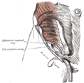
 Front of abdomen, showing surface markings for arteries and inguinal canal. (Inguinal canal is tube at lower left.) | |
 The scrotum. On the left side, the cavity of the tunica vaginalis has been opened; on the right side, only the layers superficial to the cremaster have been removed. (Right inguinal canal visible at upper left.) | |
| Details | |
| Identifiers | |
| Latin | canalis inguinalis |
| MeSH | D007264 |
| TA98 | A04.5.01.026 |
| TA2 | 2381 |
| FMA | 19928 |
| Anatomical terminology | |
The inguinal canal is a passage in the
Structure
The inguinal canals are situated just above the medial half of the inguinal ligament. The canals are approximately 4 to 6 cm long,[1] angled anteroinferiorly and medially. In males, its diameter is normally 2 cm (±1 cm in standard deviation) at the deep inguinal ring.[2][notes 1]
A first-order approximation is to visualize each canal as a cylinder.[3]
Walls
To help define the boundaries, these canals are often further approximated as boxes with six sides. Not including the two rings, the remaining four sides are usually called the "anterior wall", "inferior wall ("floor")", "superior wall ("roof")", and "posterior wall".[4] These consist of the following:
| superior wall (roof): Medial crus of aponeurosis of external oblique Musculoaponeurotic arches of internal oblique and transverse abdominal Transversalis fascia conjoint tendon | ||
| anterior wall: superficial inguinal ring (medial third of canal only)[6] |
(inguinal canal) | posterior wall: deep inguinal ring (lateral third of canal only)[6]
|
| inferior wall (floor): inguinal ligament lacunar ligament (medial third of canal only)[6] iliopubic tract (lateral third of canal only)[5] |
Deep inguinal ring
The deep inguinal ring (internal or deep abdominal ring, abdominal inguinal ring, internal inguinal ring, annulus abdominalis) is the entrance to the inguinal canal.
Location
The surface marking of the deep inguinal ring is classically described as half an inch above the midpoint of the inguinal ligament.[7]
However, the surface anatomy of the point is disputed. In a recent study,[8] it was found to be in a region between the mid-inguinal point (situated midway between the anterior superior iliac spine and the pubic symphysis) and the midpoint of the inguinal ligament (i.e. midway between the anterior superior iliac spine and the pubic tubercle). Traditionally, either one of these two sites was claimed as its location. However, this claim is based upon the study's dissection of 52 cadavers, and may not reflect the live in vivo anatomy.
Some sources state that it is at the layer of the transversalis fascia.[9]
Description
The deep inguinal ring is an opening in the
From its circumference, a thin funnel-shaped membrane, the
Superficial inguinal ring

The superficial inguinal ring (subcutaneous inguinal ring or external inguinal ring) is an anatomical structure in the anterior wall of the mammalian
It is found within the aponeurosis of the external oblique, immediately above the pubic crest, 1 centimeter above and superolateral to the pubic tubercle. It has the following boundaries—medial crura by pubic crest, lateral crura by pubic tubercle and inferiorly by inguinal ligament.[9]
Contents
The structures which pass through the canals differ between males and females:
- in males: the spermatic cord[12] and its coverings, and the ilioinguinal nerve.
- in females: the round ligament of the uterus, and the ilioinguinal nerve.
The classic description of the contents of the spermatic cords in the male are:
3 arteries: artery to vas deferens (or ductus deferens), testicular artery, cremasteric artery;
3 fascial layers: external spermatic, cremasteric, and internal spermatic fascia;
3 other structures: pampiniform plexus, vas deferens (ductus deferens), testicular lymphatics;
3 nerves: genital branch of the genitofemoral nerve (L1/2), sympathetic and visceral afferent fibres, ilioinguinal nerve (N.B. outside spermatic cord but travels next to it)
Note that the
Development
In males
During development, each
Clinical significance
Abdominal contents (potentially including intestine) can be abnormally displaced from the abdominal cavity. Where these contents exit through the inguinal canal, having passed through the
A hernia that exits the abdominal cavity directly through the deep layers of the abdominal wall, thereby bypassing the inguinal canal, is known as a
In males with strong presentation of the cremasteric reflex, the testes can—during supine sexual activity or manual manipulation—partially or fully retract into the inguinal canal for a short period of time. In juveniles and adults with inguinal injury, retraction can be prolonged and potentially lead to overheating-related infertility.[13]
The superficial ring is palpable[14] under normal conditions. It becomes dilated in a condition called athletic pubalgia. Abdominal contents may protrude through the ring in inguinal hernia.
Thus lymphatic spread from a testicular tumour is to the para-aortic nodes first, and not the inguinal nodes.
Additional images
-
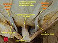
 The spermatic cord in the inguinal canal
 -
 Inguinal fossae
 -
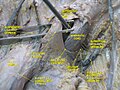
 The abdominal inguinal ring
 -
 The relations of the femoral and abdominal inguinal rings, seen from within the abdomen. Right side.
 -
 Diagram of anmedianview from the left)
 -
 Superficial inguinal ring
 -
 Anterior abdominal wall. Intermediate dissection. Anterior view.


See also
- Superficial inguinal ring
- Inguinal hernia
- Tucking
Notes
- ^ The diameter has been estimated to be ±2.2cm ±1.08cm in Africans, and 2.1 cm ±0.41cm in Europeans.
References
- PMID 29261933. Retrieved 18 June 2023.
- PMID 29643962.
- ^ "Gross Anatomy Image". Archived from the original on 2007-11-11. Retrieved 2007-11-20.
- ISBN 0-443-06612-4.
- ^ ISBN 0-7817-3639-0.
- ^ ISBN 0-7817-4255-2.
- ISBN 0-443-07168-3.
- S2CID 30726776.
- ^ ISBN 0-7817-5309-0.
- ISBN 978-0-7295-3752-0.
- ^ James Harmon, M.D., Ph.D., Lecture 13. Human Gross Anatomy. University of Minnesota. September 4, 2008.
- ^ "Anatomy Tables - Inguinal Region". Archived from the original on 2007-11-21. Retrieved 2007-11-20.
- ^ Mayo Clinic Staff. "Retractile testicle". Mayo Clinic. Mayo Foundation for Medical Education and Research. Retrieved 10 February 2018.
- ^ Moore & Agur, Essential Clinical Anatomy (2007)
- Adam Mitchell; Drake, Richard; Gray, Henry David; Wayne Vogl (2010). Gray's anatomy for students. Elsevier/Churchill Livingstone. pp. 286. ISBN 0-443-06612-4.
External links
- Anatomy photo:36:01-0102 at the SUNY Downstate Medical Center
- Anatomy figure: 36:01-03 at Human Anatomy Online, SUNY Downstate Medical Center - "The inguinal canal and derivation of the layers of the spermatic cord."
- Anatomy image:7362 at the SUNY Downstate Medical Center
- Anatomy photo:35:09-0101 at the SUNY Downstate Medical Center - "Anterior Abdominal Wall: Borders of the Superficial Inguinal Ring"
- Anatomy figure: 36:01-13 at Human Anatomy Online, SUNY Downstate Medical Center - "The inguinal canal and derivation of the layers of the spermatic cord."
- Atlas image: abdo_wall63 at the University of Michigan Health System - "The Male & Female Inguinal Canal"
- Diagram at nurseminerva.co.uk
- inguinalregion at The Anatomy Lesson by Wesley Norman (Georgetown University)
- Atlas image: abdo_wall65 at the University of Michigan Health System - "The Coverings of the Inguinal Canal, External & Internal Oblique & Transversus Abdominis Removed"
| National | |
|---|---|
| Other | |