
Otic vesicle
| Otic vesicle | |
|---|---|
auditory pit or otic pit, otic cup, | |
| Gives rise to | Membranous labyrinth of the inner ear |
| Identifiers | |
| Latin | vesicula otica |
| TE | vesicle_by_E5.15.1.0.0.0.4 E5.15.1.0.0.0.4 |
| Anatomical terminology] | |
Otic vesicle, or auditory vesicle, consists of either of the two sac-like invaginations formed and subsequently closed off during embryonic development. It is part of the neural
The general sequence in formation of the otic vesicle is relatively conserved across
Development
The otic vesicle is derived from the cranial placode.[3] The early otic vesicle is characterized as having broad competence and can be subdivided into sensory, non-sensory, and neurogenic components. Sensory epithelial cells and neurons are derived from the proneurosensory domain. This domain can be further sub-categorized into the neurogenic sub-domain and prosensory sub-domain. Prosensory sub-domain eventually gives rise to the support cells and hair cells while the neurogenic sub-domain gives rise to the auditory neuron and vestibular neuron.
The middle part of the otic vesicle develops into the ductus and
Gene signaling
The FGF, Bmp, Wnt and Pax genes are likely to be involved in otic induction.[5] FGF and BMP signals help control patterning in the early otic vesicle. Fgf3 and Fgf10 are suggested to play a role in otic induction in mice, as were Msx genes suggested to play a role in otic vesicle formation in chicks. Pax8 is expressed during the entirety of otic vesicle formation. Other genes found in the otic vesicle across species that may play a role in patterning include Hmx, Fox, Dlx, and Gbx genes.
Other animals
Formation of the otic vesicle has been studied extensively in developmental model organisms including chicken, Xenopus, zebrafish, axolotl, and mouse.[6] The transition from the otic placode to the otic vesicle occurs during the 19th somite stage in zebrafish, Xenopus, and the chick. In the chick, invagination of the otic placode occurs passively due to the movements of the surrounding placode. The otic placode in zebrafish, on the other hand, occurs by cavitation; the ectodermal placode condenses and forms an ovoid ball directly below the embryo surface. Otic vesicle formation occurs later, during the 25-30 somite stage in mice.
Additional images
-
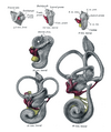 Lateral views of membranous labyrinth and acoustic complex. X 25 dia.
 -
 Median views of membranous labyrinth and acoustic complex in human embryos. X 25 dia.

