
Varicocele
| Varicocele | |
|---|---|
 | |
| Varicocele on the person's left testicle. Notice the swelling and slight enlargement of the left testicle, which is marked by an arrow. | |
| Pronunciation | |
| Specialty | Urology |
A varicocele is, in a male person, an abnormal enlargement of the
The incidence of varicocele increase with age.Signs and symptoms
Varicocele might be noticed as soft lumps, usually above the testicle and mostly on the left side of the scrotum.[5] Right-sided and bilateral varicocele does also occur. Men with varicocele can feel symptoms of pain or heaviness in their scrotum.[5] Large varicoceles present as plexus of veins and may be described as a "bag of worms".[6][7] Varicocele is sometimes discovered when investigating the cause of male infertility.[8][9]
Cause

There are three main theories as to the anatomical cause; the first has to do with the geometry of the veins, wherein the vein on the left side connects to the larger outflowing vein at a right angle, which tends to fail; the second is that testicular valves that are supposed to prevent backflow fail (venous insufficiency) leading to swelling and compression of the valveless pampiniform plexus; the third is due to excessive pressure in upstream veins, created by nutcracker syndrome.[10]
Pathophysiology
Often the greatest concern with respect to varicocele is its effect on male fertility. The relationship between varicocele and infertility is unclear. Some men with the condition are fertile, some have sperm that are normal in shape and move normally but are compromised in function, and some have sperm with abnormal shapes or that do not move well.[10] Theories as to how varicocele affects sperm function include damage via excess heat caused by the blood pooling and oxidative stress on sperm.[4][10][11][12]
Tobacco smoking and mutations in the gene expressing glutathione S-transferase Mu 1 both put men at risk for infertility; these factors may also exacerbate the risk that varicocele will affect fertility.[10]
Diagnosis
Following discovery of the sign of swelling comprising a mass, varicocele can be confirmed with scrotal ultrasound, which will show dilation of the vessels of the pampiniform plexus to be greater than 2,3 mm.[13]
-
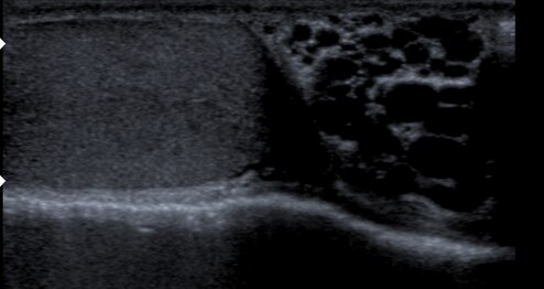 Varicocele in ultrasound (left: testicle)
 -
 Varicocele


Criteria
A universally accepted system for categorizing varicoceles does not exist, but according to the classification criteria established in 1970 by Dubin and Amelar, most commonly varicoceles are clinically assessed and categorized into three grades as follows:[14][15]
- Grade 1 Varicocele: Characterized by the palpability of the varicocele only when the individual is in a standing position during the Valsalva maneuver.
- Grade 2 Varicocele: The varicocele is palpable not only during the Valsalva maneuver but also at rest while standing.
- Grade 3 Varicocele: The most severe grade, where the varicocele is not only palpable but also visible through the scrotal skin, without any additional maneuvers.
In the Sarteschi (1993) classification system, varicoceles are categorized into five grades:[16]
- Grade I: Reflux occurs solely at the groin level during the Valsalva maneuver, without evident scrotal deformation or testicular atrophy.
- Grade II: Reflux is limited to the proximal segment of the pampiniform plexus during the Valsalva maneuver, without scrotal deformation or testicular atrophy.
- Grade III: Reflux occurs in the distal vessels located at the lower scrotum exclusively during the Valsalva maneuver, and there is no scrotal deformation or testicular atrophy.
- Grade IV: Spontaneous reverse blood flow is present and intensifies during the Valsalva maneuver, resulting in scrotal deformation and the potential for testicular atrophy.
- Grade V: Resting reflux is evident within the dilated pampiniform plexus, possibly escalating during the Valsalva maneuver, and is consistently accompanied by testicular atrophy.
Imaging
Manual examination of scrotum is required for proper interpretation of ultrasound images. During ultrasound examination, diameters of veins in pampiniform plexus are measured and regurgitation is measured. The subject is then instructed to stand up and Valsalva maneuver is performed. The diameter is then measured and changes in blood flow direction is recorded to assess any regurgitation.[17]
Treatment

The two most common surgical approaches are
Prognosis
Whether having varicocele surgery or embolization improves male fertility is controversial, as good clinical data is lacking.
Epidemiology
Around 15% to 20% of all adult males, up to 35% to 40% of men who are evaluated for male infertility, and around 80% of men who are infertile due to some other cause, have varicocele.[3][4][9]
External links
References
- ^ "Varicocele". Merriam-Webster.com Dictionary. Retrieved 2016-01-21.
- ^ "Varicocele". Lexico UK English Dictionary. Oxford University Press. Archived from the original on 2020-12-04.
- ^ a b White, Wesley M.; Kim, Edward David; Mobley, Joe D (2 January 2019). "Varicocele: Epidemiology". Medscape. Retrieved 18 September 2019.
Although varicoceles appear in approximately 20% of the general male population, they are much more common in the subfertile population (40%).
- ^ PMID 26568883.
- ^ a b "Testicular lumps and swellings - Causes - NHS Choices". NHS Choices. 7 October 2014.
- PMID 31500355.
- PMID 28846314. Retrieved 20 January 2020.
- ^ "Low sperm count". NHS Choices. 2 August 2016.
- ^ PMID 21733620.
- ^ PMID 21716891.
- PMID 27132580.
- S2CID 5477034.
- ISBN 978-93-5025-260-4.)
{{cite book}}: CS1 maint: location (link - PMID 34038625.
- S2CID 5203944.
- ^ Namdev, Rupesh. "Varicocele grading on color Doppler | Radiology Reference Article | Radiopaedia.org". Radiopaedia. Retrieved 2023-10-04.
- PMID 28138407.
- PMID 23515200.
- S2CID 20329833.
- PMID 33890288.
- PMID 26168774.