
Arachnoid granulation
| Arachnoid granulation | |
|---|---|
 Diagrammatic representation of a section across the top of the skull, showing the membranes of the brain, etc. ("Arachnoid granulation" label is at top right.) | |
 Arachnoid granulations seen on autopsy, where the dura mater has been removed but the arachnoid mater is left in place. | |
| Details | |
| Identifiers | |
| Latin | granulationes arachnoideae |
| TA98 | A14.1.01.205 |
| TA2 | 5389 |
| FMA | 77760 |
| Anatomical terminology | |
Arachnoid granulations (also arachnoid villi, and pacchionian granulations or bodies) are small protrusions of the
The largest granulations lie along the
Function
Diffusion across the arachnoid granulations into the superior sagittal sinus returns CSF to the venous circulation.[1]
The arachnoid granulations act as one-way
The importance of arachnoid granulations for the drainage of CSF is controversial. By some accounts, a large portion (perhaps the majority) of CSF is drained through lymphatics associated with extracranial segments of the cranial nerves. A large proportion of CSF is believed to leave the cranial vault through the axons of CN I (olfactory nerve) through their extension through the cribriform plate.[3]
On the inner surface of cranial bones, small pits called granular fovea are produced by arachnoid granulations.[4][5]
Eponym
Occasionally, they are referred to by their old name: Pacchioni's granulations or pacchionian bodies, named after Italian anatomist Antonio Pacchioni.[6]
References
- ISBN 978-0-321-88332-2.
- PMID 33074399.
- PMID 31063132.
- ^ Linden Forest Edwards (1934). Anatomy for physical education, descriptive and applied. P. Blakiston's son & co., inc. p. 80. Retrieved 23 June 2012.
- ^ Sir Henry Morris (1921). Morris's human anatomy. P. Blakiston's son & Company. p. 953. Retrieved 23 June 2012.
- Who Named It?
Additional images
-
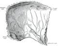 Left parietal bone. Inner surface.
 -
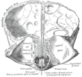 Frontal bone. Inner surface.
 -
 CT angiography showing an arachnoid granulation in the right transverse sinus
 -
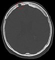 Non-contrast CT scan of the head showing an arachnoid granulation


