
Transverse sinuses
| Transverse sinuses | |
|---|---|
sigmoid sinuses | |
| Identifiers | |
| Latin | sinus transversus durae matris |
| MeSH | D054064 |
| TA98 | A12.3.05.102 |
| TA2 | 4849 |
| FMA | 50763 |
| Anatomical terminology] | |
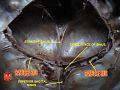
The transverse sinuses (left and right lateral sinuses), within the human head, are two areas beneath the brain which allow blood to drain from the back of the head. They run laterally in a
Structure
The transverse sinuses are of large size and begin at the internal occipital protuberance; one, generally the right, being the direct continuation of the superior sagittal sinus, the other of the straight sinus.
Each transverse sinus passes lateral and forward, describing a slight curve with its convexity upward, to the base of the
In its course it rests upon the
The transverse sinuses are frequently of unequal size, with the one formed by the superior sagittal sinus being the larger; they increase in size as they proceed, from back to center.
On transverse section, the horizontal portion exhibits a prismatic form, the curved portion has a semicylindrical form.
They receive the blood from the superior petrosal sinuses at the base of the petrous portion of the temporal bone; they communicate with the veins of the
The petrosquamous sinus, when present, runs backward along the junction of the squama and petrous portion of the temporal, and opens into the transverse sinus.
Additional images
-
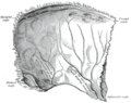 Left parietal bone. Inner surface.
 -
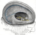 Dura mater and its processes exposed by removing part of the right half of the skull, and the brain
 -
 The sinuses at the base of the skull
 -
 Horizontal section through left ear; upper half of section
 -
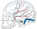 Relations of the brain and middle meningeal artery to the surface of the skull
 -
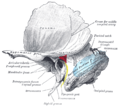 Left temporal bone showing surface markings for the tympanic antrum (red), transverse sinus (blue), and facial nerve (yellow)
 -
 Transverse sinuses
 -
 Transverse sinuses


See also
References
 This article incorporates text in the public domain from page 657 of the 20th edition of Gray's Anatomy (1918)
External links
- Cerebral Venous Sinuses at neuroangio.org