
Search results
There is a page named "Clonal deletion" on Wikipedia
- immunology, clonal deletion is the process of removing T and B lymphocytes from the immune system repertoire. The process of clonal deletion helps prevent...14 KB (1,642 words) - 00:20, 7 January 2025
- pre-existing clones of lymphocytes, which expand in response to specific antigen (process called "clonal selection"). This specific clonal army then combats...16 KB (1,936 words) - 02:38, 9 April 2025
- anemone, also called clonal anemone Vegetative cloning, a form of asexual reproduction in plants Clonal reproduction Clonal deletion, a process by which...837 bytes (140 words) - 22:12, 12 January 2022
- Peripheral tolerance (section Peripheral deletion)food antigens and allergens. Self reactive cells are subject to clonal deletion or clonal diversion. Both processes of peripheral tolerance control the...24 KB (2,982 words) - 00:30, 23 May 2025
 strongly to self-antigen, then the B cell undergoes one of four fates: clonal deletion, receptor editing, anergy, or ignorance (B cell ignores signal and...32 KB (3,744 words) - 10:38, 22 June 2025
- tolerance. Just as in T cells, clonal deletion and clonal anergy can physically eliminate autoreactive B cell clones. Receptor editing is another mechanism...28 KB (4,123 words) - 10:55, 24 May 2025
 ability to deal with these autoreactive clones via mediation of the processes of central tolerance, namely clonal deletion or T regulatory cells selection, respectively...29 KB (3,544 words) - 18:20, 28 October 2024
- bind a self peptide, then they are signaled to apoptose (process of clonal deletion). The thymic epithelial cells display self antigen to the T cells to...32 KB (4,109 words) - 03:42, 26 May 2025
 receptor. Both activated T cells and B cells express Fas and undergo clonal deletion by the AICD mechanism. Activated T cells that express both Fas and...4 KB (475 words) - 17:58, 21 April 2025
- processes of central tolerance take place in thymic medulla, namely clonal deletion (recessive tolerance) and T Regulatory cells selection (dominant tolerance)...16 KB (1,927 words) - 18:59, 28 June 2025
 Three hypotheses have gained widespread attention among immunologists: Clonal deletion theory, proposed by Burnet, according to which self-reactive lymphoid...51 KB (5,903 words) - 01:42, 1 July 2025
 In mice models, most mother's fetal-specific CD8+ T cells undergo clonal deletion and express low levels of chemokine receptors and ligands – this prevents...43 KB (5,119 words) - 09:55, 30 June 2025
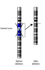 In genetics, a deletion (also called gene deletion, deficiency, or deletion mutation) (sign: Δ) is a mutation (a genetic aberration) in which a part of...14 KB (1,537 words) - 13:41, 10 March 2025
- is allowed to proliferate or is killed off through a process called clonal deletion. Normally, the immune system is able to recognize and ignore the body's...18 KB (1,555 words) - 19:01, 1 March 2024
- 16p11.2 deletion syndrome is a rare genetic condition caused by microdeletion on the short arm of chromosome 16. Most affected individuals experience global...14 KB (1,064 words) - 23:48, 7 July 2024
 Yin L, Zheng X, et al. (July 2008). "Autoreactive T cells escape clonal deletion in the thymus by a CD24-dependent pathway". Journal of Immunology....12 KB (1,412 words) - 07:52, 30 June 2025
- were the first to propose the deletion of self-reactive lymphocytes to establish tolerance, now termed clonal deletion. Burnet and Medawar were ultimately...50 KB (6,091 words) - 17:55, 21 April 2025
- Sprent, Jonathan, and Susan R. Webb. "Intrathymic and extrathymic clonal deletion of T cells." Current opinion in immunology 7.2 (1995): 196-205. Crotty...24 KB (2,961 words) - 23:06, 22 March 2025
- activity is selective but is not T-cell receptor mediated. Both clonal anergy and clonal deletion have been shown to operate in vetoed T cells. The veto cell...18 KB (2,148 words) - 21:10, 19 December 2023
- abrogated by thymus-dependent dextran conjugates: evidence against clonal deletion as the mechanism of tolerance induction". Scandinavian Journal of Immunology...7 KB (717 words) - 14:51, 23 May 2025


- interactions. There is an association of missense mutations and small in frame deletions with a higher mean age at onset of renal disease and with absence of neurologic
- before the changes are completed. Also, in step 530, the creation and deletion of snapshots, described below, are performed because it is the only point
- low, up to ~40%; median OS is ~8 months 1993 PMID 7693038, 1993 — "Common clonal origin of chronic lymphocytic leukemia and high-grade lymphoma of Richter's