
Great auricular nerve
| Great auricular nerve | |
|---|---|
 | |
 Plan of the cervical plexus. (Great auricular labeled at top center.) | |
| Details | |
| From | cervical plexus (C2-C3) |
| Innervates | sensation of inferior part of auricle and parotid region of the face. |
| Identifiers | |
| Latin | nervus auricularis magnus |
| TA98 | A14.2.02.018 |
| TA2 | 6385 |
| FMA | 6872 |
| Anatomical terms of neuroanatomy | |
The great auricular nerve is a cutaneous (sensory) nerve of the head. It originates from the second and third cervical (spinal) nerves (C2-C3) of the cervical plexus. It provides sensory innervation to the skin over the parotid gland and the mastoid process, parts of the outer ear, and to the parotid gland and its fascia.
Pain resulting from parotitis is caused by an impingement on the great auricular nerve.
Structure
The great auricular nerve is the largest of the ascending branches of the cervical plexus.[1]
Origin
It arises from the second and third cervical (spinal) nerves (C2-C3),[1] with the predominant contribution coming from C2.[2]
Course and relations
The great auricular nerve is a large trunk that ascends almost vertically over the sternocleidomastoid.[2] It winds around the posterior border of the sternocleidomastoid muscle, then perforates the deep fascia before ascending alongside the external jugular vein upon that sternocleidomastoid muscle beneath the platysma muscle to the parotid gland.[1] Upon reaching the parotid gland, it divides into an anterior branch and a posterior branch.[1]
Branches
Anterior branch
The anterior branch (or facial branch[citation needed]) is distributed to the skin of the face over the parotid gland.[1]
It communicates with the facial nerve (CN VII) inside the parotid gland.[1]
Posterior branch
The posterior branch (or mastoid branch[
The posterior branch communicates with the
Distribution
The great auricular nerve is distributed to the skin of the face over the angle of the mandible[2] and parotid gland (via anterior branch[1]),[1][2] skin over of the mastoid region[2] (i.e. skin over the mastoid process[1]) (via posterior branch[1]), parts of the auricle (posterior branch[1]),[1][2] and the parotid gland and parotid fascia.[2]
Clinical significance
The great auricular nerve may be damaged during surgery on the parotid gland, reducing sensation to the face.[3]
Pain resulting from parotitis is caused by an impingement on the great auricular nerve.[citation needed]
The intermingling of the great auricular nerve and the facial nerve (CN VII) is thought to be responsible for the pathogenesis of Frey's syndrome following parotidectomy.[1]
Additional images
-
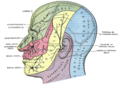 Dermatome distribution of the trigeminal nerve, also showing the sensory distribution of the great auricular, lesser occipital, and greater occipital nerves.
 -
 Side of neck, showing chief surface markings.


References
- ^ OCLC 1201341621.)
{{cite book}}: CS1 maint: location missing publisher (link - ^ ISBN 978-0-7295-3752-0.
- PMID 2597656.
External links
- Diagram at aapmr.org
- Anatomy figure: 25:03-03 at Human Anatomy Online, SUNY Downstate Medical Center - "Diagram of the cervical plexus."