
External jugular vein
| External jugular vein | |
|---|---|
posterior facial vein, posterior auricular vein, retromandibular vein, anterior jugular vein, transverse cervical vein, suprascapular vein | |
| Drains to | subclavian vein |
| Identifiers | |
| Latin | vena jugularis externa |
| TA98 | A12.3.05.045 |
| TA2 | 4956 |
| FMA | 13110 |
| Anatomical terminology] | |
The external jugular vein receives the greater part of the blood from the exterior of the
Structure
It commences in the substance of the
In its course it crosses the sternocleidomastoideus obliquely, and in the
It is separated from the sternocleidomastoideus by the investing layer of the deep cervical fascia, and is covered by the
Valves
It is provided with two pairs of valves, the lower pair being placed at its entrance into the subclavian vein, the upper in most cases about 4 cm above the clavicle. The portion of vein between the two sets of valves is often dilated, and is termed the sinus.
These valves do not prevent the regurgitation of the blood, or the passage of injection from below upward.[2]
Variation
The external jugular vein varies in size, bearing an inverse proportion to the other veins of the neck, it is occasionally double.[3]
Function
This vein receives the
The external jugular vein drains into the subclavian vein lateral to the junction of the subclavian vein and the internal jugular vein.
Clinical significance
The external jugular is a large vein used in
Additional images
-
 Veins of the thoracic and abdominal regions
 -
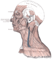 Muscles of the head, face, and neck.
 -
 Section of the neck at about the level of the sixth cervical vertebra.
 -
 The venæ cavæ and azygos veins, with their tributaries.


See also
- Chronic cerebrospinal venous insufficiency
- Jugular vein
References
![]() This article incorporates text in the public domain from page 646 of the 20th edition of Gray's Anatomy (1918)
This article incorporates text in the public domain from page 646 of the 20th edition of Gray's Anatomy (1918)
- ^ [Standring, S., & Gray, H. (2016). Grays anatomy: the anatomical basis of clinical practice. Philadelphia: Elsevier. p.414]
- ^ Gray's Anatomy of the Human Body
- . Retrieved September 1, 2015.
- ^ http://pehsc.org/wp-content/uploads/2015/08/EMS-Provider-Scope-of-Practice-08-28-15.pdf [bare URL PDF]