Hypogastric nerve
| Hypogastric nerve | |
|---|---|
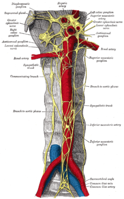
 Autonomic plexuses and ganglia on the abdominal aorta. (Hypogastric nerves visible at the bottom of the image but not labeled.) | |
| Details | |
| Identifiers | |
| Latin | nervus hypogastricus |
| TA98 | A14.3.03.047 |
| TA2 | 6714 |
| FMA | 77596 |
| Anatomical terms of neuroanatomy | |
The hypogastric nerves (one on each side) are the continuation of the superior hypogastric plexus that descend into the pelvis anterior the sacrum and become the inferior hypogastric plexuses on either side of pelvic organs. The hypogastric nerves serve as a pathway for autonomic fibers to communicate between the lower abdomen and pelvis.
Structure
The hypogastric nerves begin where the superior hypogastric plexus splits into a right and left hypogastric nerves. The hypogastric nerves continue inferiorly on their corresponding side of the body, where they descends into the pelvis to form the inferior hypogastric plexuses.[1]
The hypogastric nerves likely contain three nerve fibers types:[2]
- Preganglionic and postganglionic sympathetic fibers descend from the superior hypogastric plexus from lumbar splanchnic nerves (from the sympathetic trunk at levels L1-L2). Sympathetic fibers are the most numerous fibers in the hypogastric nerves.[2]
- Preganglionic parasympathetic fibers that originate from pelvic splanchnic nerves (sacral spinal nerves, S2-S4) ascend from the inferior hypogastric plexuses into hypogastric nerves.[2][3]
- Visceral sensory fibers that project to the lumbar spinal cord.[2]
Clinical significance
The hypogastric nerve may be blocked for a local anaesthetic.[4] This endangers the nearby common iliac artery and common iliac vein.[4]
See also
References
- PMID 32427364.
- ^ S2CID 23908051.
- OCLC 1202943188.
- ^ ISBN 978-0-7216-0334-6, retrieved 2021-02-06
External links
- Anatomy photo:40:09-0203 at the SUNY Downstate Medical Center - "Posterior Abdominal Wall: The Abdominal Aorta and Paraaortic Nerve Plexus"
- Autonomics of the Pelvis - Page 5 of 12 anatomy module at med.umich.edu
- https://web.archive.org/web/20060709234151/http://www.downstate.edu/ginzler-painmanagement/ginzler-painmanagement.htm
