
Vas deferens
This article needs additional citations for verification. (March 2013) |
| Vas deferens | |
|---|---|
artery of the ductus deferens | |
| Lymph | External iliac lymph nodes, internal iliac lymph nodes |
| Identifiers | |
| Latin | vas deferens (plural: vasa deferentia), ductus deferens (plural: ductus deferentes) |
| MeSH | D014649 |
| TA98 | A09.3.05.001 |
| TA2 | 3621 |
| FMA | 19234 |
| Anatomical terminology] | |

The vas deferens (pl.: vasa deferentia), with the more modern name ductus deferens (pl.: ductūs deferentes), is part of the male
Etymology
Vas deferens is Latin, meaning "carrying-away vessel" while ductus deferens, also Latin, means "carrying-away duct".[1]
Structure
The human vas deferens measures 30–35 cm in length, and 2–3 mm in diameter.
Blood supply
The vasa deferentia are supplied with blood by accompanying arteries, the (
Innervation
The vas deferens receives innervation from an autonomic plexus of post-ganglionic sympathetic fibres derived from the inferior hypogastric plexus.[2]: 1297
It is innervated by a variety of
Anatomical relations
Within the spermatic cord, the vas deferens is situated posterior (and parallel to) the vessels of the spermatic cord.[2]: 1297
The vas deferens traverses the inguinal canal to reach the
Histology
The vas deferens consists of an external adventitial sheath containing blood vessels and nerves, a muscular middle layer composed of three layers of smooth muscle (with a circular muscle layer interposed between two longitudinal muscle layers), and an internal mucosal lining consisting of pseudostratified columnar epithelium (which bears the non-motile stereocilia).[2]: 1297 [9]
The vas deferens has the greatest muscle-to-lumen ratio of any hollow organ.[2]: 1297
Function
During
Clinical significance
Contraception
A
Disease
The vas deferens may be obstructed, or it may be completely absent in a condition known as congenital absence of the vas deferens (CAVD, a potential feature of cystic fibrosis), causing male infertility. Acquired obstructions can occur due to infections. To treat these causes of male infertility, sperm can be harvested by testicular sperm extraction (TESE) or microsurgical epididymal sperm aspiration (MESA).[13]
Uses in pharmacology and physiology
The vas deferens has a dense sympathetic innervation,[14] making it a useful system for studying sympathetic nerve function and for studying drugs that modify neurotransmission.[6]
It has been used:
- as a bioassay for the discovery of enkephalins, the endogenous opiates.[15]
- to demonstrate quantal transmission from sympathetic nerve terminals.[16]
- as the first direct measure of free Ca2+ concentration in a postganglionic nerve terminal.[17]
- to develop an optical method for monitoring packeted transmission (similar to quantal transmission).[18]
Other animals
Most vertebrates have some form of duct to transfer the sperm from the
In cartilaginous fishes, the part of the archinephric duct closest to the testis is coiled up to form an epididymis. Below this are a number of small glands secreting components of the seminal fluid. The final portion of the duct also receives ducts from the kidneys in most species.[19]
In amniotes, however, the archinephric duct has become a true vas deferens, and is used only for conducting sperm, never urine. As in cartilaginous fish, the upper part of the duct forms the epididymis. In many species, the vas deferens ends in a small sac for storing sperm.[19]
The only vertebrates to lack any structure resembling a vas deferens are the primitive
Additional images
-
 Male reproductive system.
 -
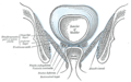 Coronal section of pelvis, showing arrangement of fasciae. Viewed from behind.
 -
 The relations of the femoral and abdominal inguinal rings, seen from within the abdomen. Right side.
 -
 The spermatic cord in the inguinal canal.
 -
 Fundus of the bladder with the vesiculae seminales.
 -
 Vertical section of bladder, penis, and urethra.
 -
 Prostate with seminal vesicles and seminal ducts, viewed from in front and above.
 -
 Prostate
 -
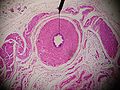 Microscopic cross section.
 -
 Testis, spermatic vessels and vas deferens
 -
 A deep dissection showing the vas deferens.


See also
- Intra vas device
- Excretory duct of seminal gland
- Vas deferens in the reproductive system of gastropods
References
- S2CID 245049526.
- ^ OCLC 1201341621.)
{{cite book}}: CS1 maint: location missing publisher (link) CS1 maint: others (link - PMID 11753468.
- S2CID 81721236.
- ^
 One or more of the preceding sentences incorporates text in the public domain from page 615 of the 20th edition of Gray's Anatomy (1918)
- ^ PMID 20074819.
- .
- S2CID 4761228.
- S2CID 7457415.
- PMID 18787034.
- ^ Mann, T (1954). The Biochemistry of Semen. London: Methuen & Co; New York: John Wiley & Sons. Retrieved November 9, 2013.
- PMID 24683020.
- PMID 11098022.
- ^ Sjöstrand, N.O. (1965). "The adrenergic innervation of the vas deferens and the accessory male genital organs". Acta Physiologica Scandinavica. 257: S1–82.
- S2CID 95411.
- S2CID 4303337.
- PMID 9279805.
- PMID 12068045.
- ^ ISBN 978-0-03-910284-5.
- ISBN 978-0-643-06257-3.
- ISBN 978-1-139-45742-2.
External links
- Anatomy photo:36:07-0301 at the SUNY Downstate Medical Center—"Inguinal Region, Scrotum and Testes: Layers of the Spermatic Cord"
- Anatomy photo:44:02-0301 at the SUNY Downstate Medical Center—"The Male Pelvis: Distribution of the Peritoneum in the Male Pelvis"
- MedicalMnemonics.com: 2424 319 [dead link]
- Cross section image: pelvis/pelvis-e12-15—Plastination Laboratory at the Medical University of Vienna
- inguinalregion at The Anatomy Lesson by Wesley Norman (Georgetown University) (testes)