
Lamina terminalis
| Lamina terminalis | |
|---|---|
 Median sagittal section of brain of human embryo of three months. (Lamina terminalis labeled at center left.) | |
 Median sagittal section of brain of human embryo of four months. (Lamina terminalis labeled at center right.) | |
| Details | |
| Identifiers | |
| Latin | lamina terminalis |
| NeuroNames | 208 |
| TA98 | A14.1.08.419 |
| TA2 | 5776 |
| FMA | 61975 |
| Anatomical terms of neuroanatomy | |
The median portion of the wall of the
vascular organ of the lamina terminalis, which regulates the osmotic concentration of the blood. The lamina terminalis is immediately anterior to the tuber cinereum; together they form the pituitary stalk
.
The lamina terminalis can be opened via
endoscopic neurosurgery in an attempt to create a path that cerebrospinal fluid can flow through when a person has hydrocephalus and when it is not possible to perform an endoscopic third ventriculostomy,[1] but the effectiveness of this technique is not certain.[2]
This is the rostral end (tip) of the neural tube (embryological central nervous system) in the early weeks of development. Failure of the lamina terminalis to close properly at this stage of development will result in anencephaly or meroencephaly.
Additional images
-
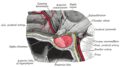
 Mesal aspect of a brain sectioned in the median sagittal plane.
 -
 Thesagittalsection.
 -
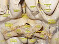 Cerebrum. Inferior view. Deep dissection


See also
References
![]() This article incorporates text in the public domain from page 816 of the 20th edition of Gray's Anatomy (1918)
This article incorporates text in the public domain from page 816 of the 20th edition of Gray's Anatomy (1918)
- PMID 20707616.
- PMID 19284236.
- ISBN 9780134320823.