
Hypothalamus
| Hypothalamus | |
|---|---|
 Location of the human hypothalamus | |
 Location of the hypothalamus (cyan) in relation to the pituitary and to the rest of the brain | |
| Details | |
| Part of | Brain |
| Identifiers | |
| Latin | hypothalamus |
| MeSH | D007031 |
| NeuroLex ID | birnlex_734 |
| TA98 | A14.1.08.401 A14.1.08.901 |
| TA2 | 5714 |
| FMA | 62008 |
| Anatomical terms of neuroanatomy | |
The hypothalamus (pl.: hypothalami; from
The hypothalamus is responsible for regulating certain
Structure
The hypothalamus is divided into four regions (preoptic, supraoptic, tuberal, mammillary) in a parasagittal plane, indicating location anterior-posterior; and three zones (periventricular, intermediate, lateral) in the coronal plane, indicating location medial-lateral.
Nuclei
The hypothalamic nuclei include the following:[11][12]
| Region | Area | Nucleus | Function[13] |
| Anterior (supraoptic) | Preoptic | Preoptic nucleus | |
| Medial | Medial preoptic nucleus |
| |
| Supraoptic nucleus |
| ||
Paraventricular nucleus |
| ||
| Anterior hypothalamic nucleus |
| ||
| Suprachiasmatic nucleus |
| ||
| Lateral | |||
Lateral nucleus |
See Lateral hypothalamus § Function – primary source of orexin neurons that project throughout the brain and spinal cord | ||
| Middle (tuberal) | Medial | Dorsomedial hypothalamic nucleus |
|
Ventromedial nucleus |
| ||
| Arcuate nucleus | |||
| Lateral | Lateral nucleus |
See Lateral hypothalamus § Function – primary source of orexin neurons that project throughout the brain and spinal cord | |
| Lateral tuberal nuclei | |||
| Posterior (mammillary) | Medial | Mammillary nuclei (part of mammillary bodies) | |
Posterior nucleus |
| ||
| Lateral | Lateral nucleus |
See Lateral hypothalamus § Function – primary source of orexin neurons that project throughout the brain and spinal cord | |
| Tuberomammillary nucleus[15] |
|
-
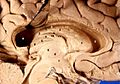
 Cross-section of the monkey hypothalamus displays two of the major hypothalamic nuclei on either side of the fluid-filled third ventricle.
 -
 Hypothalamic nuclei
 -
![Hypothalamic nuclei on one side of the hypothalamus, shown in a 3-D computer reconstruction[16]](//upload.wikimedia.org/wikipedia/commons/thumb/0/04/3D-Hypothalamus.JPG/120px-3D-Hypothalamus.JPG) Hypothalamic nuclei on one side of the hypothalamus, shown in a 3-D computer reconstruction[16]
Hypothalamic nuclei on one side of the hypothalamus, shown in a 3-D computer reconstruction[16]


![Hypothalamic nuclei on one side of the hypothalamus, shown in a 3-D computer reconstruction[16]](/File:3D-Hypothalamus.JPG)
Connections
The hypothalamus is highly interconnected with other parts of the
The hypothalamus receives many inputs from the
Most nerve fibres within the hypothalamus run in two ways (bidirectional).
- Projections to areas caudal to the hypothalamus go through the medial forebrain bundle, the mammillotegmental tract and the dorsal longitudinal fasciculus.
- Projections to areas rostral to the hypothalamus are carried by the terminal stria.
- Projections to areas of the lateral horn spinal segments T1–L2/L3) are carried by the hypothalamospinal tractand they activate the sympathetic motor pathway.
Sexual dimorphism
Several hypothalamic nuclei are
Responsiveness to ovarian steroids
Other striking functional dimorphisms are in the behavioral responses to
Male and female brains differ in the distribution of estrogen receptors, and this difference is an irreversible consequence of neonatal steroid exposure.[citation needed] Estrogen receptors (and progesterone receptors) are found mainly in neurons in the anterior and mediobasal hypothalamus, notably:
- the LHRH neurons are located, regulating dopamine responses and maternal behavior;[19]
- the periventricular nucleus where somatostatin neurons are located, regulating stress levels;[20]
- the ventromedial hypothalamuswhich regulates hunger and sexual arousal.
Development

In neonatal life, gonadal steroids influence the development of the neuroendocrine hypothalamus. For instance, they determine the ability of females to exhibit a normal reproductive cycle, and of males and females to display appropriate reproductive behaviors in adult life.
- If a female rat is injected once with testosterone in the first few days of postnatal life (during the "critical period" of sex-steroid influence), the hypothalamus is irreversibly masculinized; the adult rat will be incapable of generating an LH surge in response to estrogen (a characteristic of females), but will be capable of exhibiting male sexual behaviors (mounting a sexually receptive female).[21]
- By contrast, a male rat castrated just after birth will be feminized, and the adult will show female sexual behavior in response to estrogen (sexual receptivity, lordosis behavior).[21]
In primates, the developmental influence of
Function
Hormone release

The hypothalamus has a central
- Anterior pituitary
In the hypothalamic–adenohypophyseal axis, releasing hormones, also known as hypophysiotropic or hypothalamic hormones, are released from the median eminence, a prolongation of the hypothalamus, into the hypophyseal portal system, which carries them to the anterior pituitary where they exert their regulatory functions on the secretion of adenohypophyseal hormones.[24] These hypophysiotropic hormones are stimulated by parvocellular neurosecretory cells located in the periventricular area of the hypothalamus. After their release into the capillaries of the third ventricle, the hypophysiotropic hormones travel through what is known as the hypothalamo-pituitary portal circulation. Once they reach their destination in the anterior pituitary, these hormones bind to specific receptors located on the surface of pituitary cells. Depending on which cells are activated through this binding, the pituitary will either begin secreting or stop secreting hormones into the rest of the bloodstream.[25]
| Secreted hormone | Abbreviation | Produced by | Effect |
|---|---|---|---|
| Thyrotropin-releasing hormone (Prolactin-releasing hormone) |
TRH, TRF, or PRH | paraventricular nucleus |
Stimulate thyroid-stimulating hormone (TSH) release from anterior pituitary (primarily) Stimulate prolactin release from anterior pituitary |
| Corticotropin-releasing hormone | CRH or CRF | Parvocellular neurosecretory cells of the paraventricular nucleus | Stimulate adrenocorticotropic hormone (ACTH) release from anterior pituitary |
| Dopamine (Prolactin-inhibiting hormone) |
DA or PIH | Dopamine neurons of the arcuate nucleus | Inhibit prolactin release from anterior pituitary |
Growth-hormone-releasing hormone
|
GHRH | Neuroendocrine neurons of the Arcuate nucleus |
Stimulate growth-hormone (GH) release from anterior pituitary |
| Gonadotropin-releasing hormone | GnRH or LHRH | Neuroendocrine cells of the Preoptic area |
Stimulate follicle-stimulating hormone (FSH) release from anterior pituitary Stimulate luteinizing hormone (LH) release from anterior pituitary |
| Somatostatin[26] (growth-hormone-inhibiting hormone) |
SS, GHIH, or SRIF | Neuroendocrine cells of the Periventricular nucleus |
Inhibit growth-hormone (GH) release from anterior pituitary Inhibit (moderately) thyroid-stimulating hormone (TSH) release from anterior pituitary |
Other hormones secreted from the median eminence include vasopressin, oxytocin, and neurotensin.[27][28][29][30]
- Posterior pituitary
In the hypothalamic–pituitary–adrenal axis, neurohypophysial hormones are released from the posterior pituitary, which is actually a prolongation of the hypothalamus, into the circulation.
| Secreted hormone | Abbreviation | Produced by | Effect |
|---|---|---|---|
| Oxytocin | OXY or OXT | Magnocellular neurosecretory cells of the paraventricular nucleus and supraoptic nucleus | Lactation (letdown reflex)
|
| Vasopressin (antidiuretic hormone) |
ADH or AVP | Magnocellular and parvocellular neurosecretory cells of the paraventricular nucleus, magnocellular cells in supraoptic nucleus | Increase in the permeability to water of the cells of collecting duct in the kidney and thus allows water reabsorption and excretion of concentrated urine
|
It is also known that hypothalamic–pituitary–adrenal axis (HPA) hormones are related to certain skin diseases and skin homeostasis. There is evidence linking hyperactivity of HPA hormones to stress-related skin diseases and skin tumors.[31]
Stimulation
The hypothalamus coordinates many hormonal and behavioural circadian rhythms, complex patterns of
The hypothalamus is responsive to:
- Light: daylength and circadianand seasonal rhythms
- pheromones
- corticosteroids
- Neurally transmitted information arising in particular from the heart, enteric nervous system (of the gastrointestinal tract),[32] and the reproductive tract.[citation needed]
- Autonomicinputs
- Blood-borne stimuli, including cytokines, plasma concentrations of glucose and osmolarity etc.
- Stress
- Invading microorganisms by increasing body temperature, resetting the body's thermostat upward.
Olfactory stimuli
Olfactory stimuli are important for
Blood-borne stimuli
It is not clear how all peptides that influence hypothalamic activity gain the necessary access. In the case of prolactin and leptin, there is evidence of active uptake at the choroid plexus from the blood into the cerebrospinal fluid (CSF). Some pituitary hormones have a negative feedback influence upon hypothalamic secretion; for example, growth hormone feeds back on the hypothalamus, but how it enters the brain is not clear. There is also evidence for central actions of prolactin.[citation needed]
Findings have suggested that
The hypothalamus functions as a type of thermostat for the body.[34] It sets a desired body temperature, and stimulates either heat production and retention to raise the blood temperature to a higher setting or sweating and vasodilation to cool the blood to a lower temperature. All fevers result from a raised setting in the hypothalamus; elevated body temperatures due to any other cause are classified as hyperthermia.[34] Rarely, direct damage to the hypothalamus, such as from a stroke, will cause a fever; this is sometimes called a hypothalamic fever. However, it is more common for such damage to cause abnormally low body temperatures.[34]
Steroids
The hypothalamus contains neurons that react strongly to steroids and
Neural
Cardiovascular stimuli are carried by the vagus nerve. The vagus also conveys a variety of visceral information, including for instance signals arising from gastric distension or emptying, to suppress or promote feeding, by signalling the release of leptin or gastrin, respectively. Again this information reaches the hypothalamus via relays in the brainstem.
In addition hypothalamic function is responsive to—and regulated by—levels of all three classical
Control of food intake
| Peptides that increase feeding behavior |
Peptides that decrease feeding behavior |
|---|---|
| Ghrelin | Leptin |
| Neuropeptide Y | (α,β,γ)-Melanocyte-stimulating hormones |
| Agouti-related peptide | Cocaine- and amphetamine-regulated transcript peptides |
| Orexins (A,B) | Corticotropin-releasing hormone |
| Melanin-concentrating hormone | Cholecystokinin |
| Galanin | Insulin |
Glucagon-like peptide 1
|
The extreme
There are different hypotheses related to this regulation:[36]
- Lipostatic hypothesis: This hypothesis holds that acts on the hypothalamus to decrease food intake and increase energy output.
- Gutpeptide hypothesis: gastrointestinal hormones like Grp, glucagons, CCK and others claimed to inhibit food intake. The food entering the gastrointestinal tract triggers the release of these hormones, which act on the brain to produce satiety. The brain contains both CCK-A and CCK-B receptors.
- Glucostatic hypothesis: The activity of the satiety center in the ventromedial nuclei is probably governed by the 2-deoxyglucosetherefore decreasing glucose utilization in cells.
- Thermostatic hypothesis: According to this hypothesis, a decrease in body temperature below a given set-point stimulates appetite, whereas an increase above the set-point inhibits appetite.
Fear processing
The medial zone of hypothalamus is part of a circuitry that controls motivated behaviors, like defensive behaviors.
- Antipredatory defensive behavior
Exposure to a predator (such as a cat) elicits defensive behaviors in laboratory rodents, even when the animal has never been exposed to a cat.
- Social defeat
Likewise, the hypothalamus has a role in social defeat: Nuclei in medial zone are also mobilized during an encounter with an aggressive conspecific. The defeated animal has an increase in Fos levels in sexually dimorphic structures, such as the medial pre-optic nucleus, the ventrolateral part of ventromedial nucleus, and the ventral premammilary nucleus.[6] Such structures are important in other social behaviors, such as sexual and aggressive behaviors. Moreover, the premammillary nucleus also is mobilized, the dorsomedial part but not the ventrolateral part.[6] Lesions in this nucleus abolish passive defensive behavior, like freezing and the "on-the-back" posture.[6]
Learning arbitrator
Recent research has questioned whether the lateral hypothalamus's role is only restricted to initiating and stopping innate behaviors and argued it learns about food-related cues. Specifically that it opposes learning about information what is neutral or distant to food. According this view, the lateral hypothalamus is "a unique arbitrator of learning capable of shifting behavior toward or away from important events".[44]
Additional images
-

-
 Human brain left dissected midsagittal view
 -
 Location of the hypothalamus


See also
- ventrolateral preoptic nucleus
- periventricular nucleus
- Copeptin
- Hypothalamic–pituitary–adrenal axis (HPA axis)
- Hypothalamic–pituitary–gonadal axis (HPG axis)
- Hypothalamic–pituitary–thyroid axis (HPT axis)
- Incertohypothalamic pathway
- Neuroendocrinology
- Neuroscience of sleep
References
- ^ Boeree CG. "The Emotional Nervous System". General Psycholoty. Retrieved 18 April 2016.
- PMID 33910896.
- PMID 26365177.
- ^ "NCI Dictionary of Cancer Terms". National Cancer Institute.
- S2CID 1793658.
- ^ PMID 19273843.
- ISBN 9788131229811.
- ISBN 978-93-5025-383-0.
- PMID 8277003.
- ISBN 978-1437703245.
- ^ "Enlarged view of the hypothalamus". psycheducation.org. Jim Phelps. Archived from the original on 15 December 2005. Retrieved 7 February 2020.
- The University of Texas at Dallas. Retrieved 7 February 2020.
- ISBN 978-1416045748.
- PMID 19776281.
- ISBN 9780071481274.
Within the brain, histamine is synthesized exclusively by neurons with their cell bodies in the tuberomammillary nucleus (TMN) that lies within the posterior hypothalamus. There are approximately 64000 histaminergic neurons per side in humans. These cells project throughout the brain and spinal cord. Areas that receive especially dense projections include the cerebral cortex, hippocampus, neostriatum, nucleus accumbens, amygdala, and hypothalamus. ... While the best characterized function of the histamine system in the brain is regulation of sleep and arousal, histamine is also involved in learning and memory ... It also appears that histamine is involved in the regulation of feeding and energy balance.
- ^ Brain Research Bulletin 35:323–327, 1994
- ^ PMID 2606795.
- PMID 25987976.
- PMID 25938107.
- S2CID 31026141.
- ^ PMID 22396398.
- PMID 16410296.
- ^ Bowen R. "Overview of Hypothalamic and Pituitary Hormones". Retrieved 5 October 2014.
- ISBN 978-0-07-139140-5.
- ISBN 978-0-7817-7817-6.
- PMID 20149677.
- PMID 3968510.
- PMID 10719209.
- PMID 8367038.
- S2CID 17941978.
- ^ Jung Eun Kim; Baik Kee Cho; Dae Ho Cho; Hyun Jeong Park (2013). "Expression of Hypothalamic–Pituitary–Adrenal Axis in Common Skin Diseases: Evidence of its Association with Stress-related Disease Activity". National Research Foundation of Korea. Retrieved 4 March 2014.
- PMID 21750565.
- S2CID 33268046.
- ^ ISBN 978-0-07-146633-2.
- ISBN 9780071481274.
- PMID 178168.
- S2CID 10167219.
- S2CID 12303256.
- S2CID 8063630.
- ^ S2CID 10073236.
- ^ S2CID 16406187.
- PMID 1279669.
- S2CID 24690642.
- PMID 37758590.
Further reading
- de Vries GJ, Södersten P (May 2009). "Sex differences in the brain: the relation between structure and function". Hormones and Behavior. 55 (5): 589–96. PMID 19446075.
External links
- Stained brain slice images which include the "Hypothalamus" at the BrainMaps project
- The Hypothalamus and Pituitary at endotexts.org
- NIF Search - Hypothalamus via the Neuroscience Information Framework
- Space-filling and cross-sectional diagrams of hypothalamic nuclei: right hypothalamus, anterior, tubular, posterior.
| National | |
|---|---|
| Other | |